. 2000 Mar;105(5):673-81.
doi: 10.1172/JCI5826.

Molecular basis of variant pseudo-hurler polydystrophy (mucolipidosis IIIC)

Affiliations

Molecular basis of variant pseudo-hurler polydystrophy (mucolipidosis IIIC)

A Raas-Rothschild et al. J Clin Invest. 2000 Mar.

Abstract

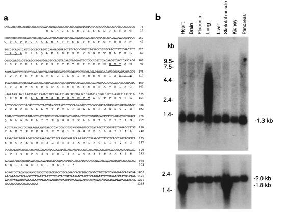
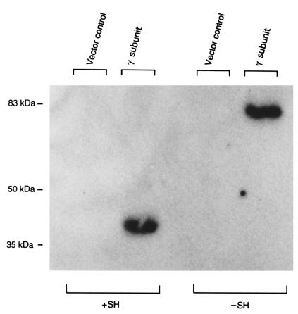
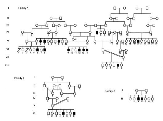
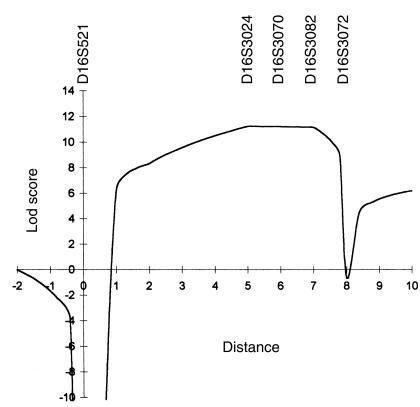
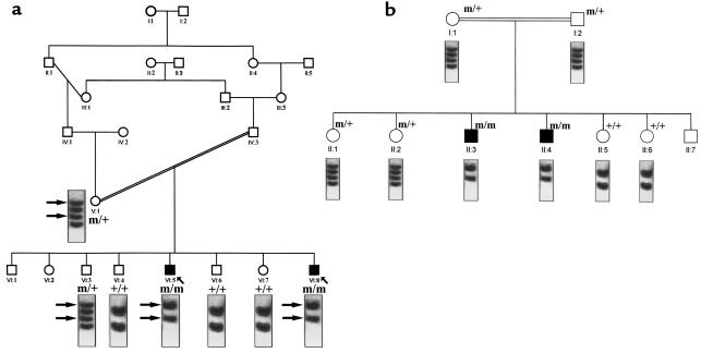
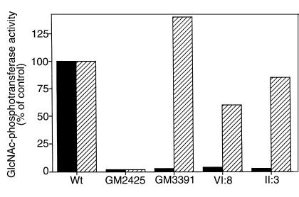
Mucolipidosis IIIC, or variant pseudo-Hurler polydystrophy, is an autosomal recessive disease of lysosomal hydrolase trafficking. Unlike the related diseases, mucolipidosis II and IIIA, the enzyme affected in mucolipidosis IIIC (N-Acetylglucosamine-1-phosphotransferase [GlcNAc-phosphotransferase]) retains full transferase activity on synthetic substrates but lacks activity on lysosomal hydrolases. Bovine GlcNAc-phosphotransferase has recently been isolated as a multisubunit enzyme with the subunit structure alpha(2)beta(2)gamma(2). We cloned the cDNA for the human gamma-subunit and localized its gene to chromosome 16p. We also showed, in a large multiplex Druze family that exhibits this disorder, that MLIIIC also maps to this chromosomal region. Sequence analysis of the gamma-subunit cDNA in patients from 3 families identified a frameshift mutation, in codon 167 of the gamma subunit, that segregated with the disease, indicating MLIIIC results from mutations in the phosphotransferase gamma-subunit gene. This is to our knowledge the first description of the molecular basis for a human mucolipidosis and suggests that the gamma subunit functions in lysosomal hydrolase recognition.


Figures

Figure 1
Figure 1
(a) Nucleotide and deduced amino acid sequence of the human GlcNAc-phosphotransferase γ-subunit cDNA. The predicted protein sequence is shown below the DNA sequence. The putative signal peptide is indicated by the dotted underline. Sequences homologous to the bovine γ subunit are indicated by single underline. Two potential sites for N-linked glycosylation are indicated by double underline. (b) Northern blot analysis of GlcNAc-phosphotransferase γ subunit. The γ-subunit cDNA was random labeled and hybridized to a human multiple tissue Northern blot (CLONTECH); each lane contained 2 μg of poly(A)+ RNA from the indicated tissue. The filter was washed at 65°C in 0.1 × SSC, 0.1% SDS, and exposed for 18 hours. In a, transcripts of 1.3 kb are identified in all tissues with additional larger transcripts identified in lung; in b, after hybridization, the blot was stripped and rehybridized with a probe for human β-actin mRNA as a control for loading.
Figure 2
Figure 2
Expression of human GlcNAc-phosphotransferase γ subunit in COS cells. Membrane fractions of COS cells transfected with either the epitope-tagged γ-subunit cDNA or a vector control were analyzed by immunoblot. The samples were analyzed by 7.5% SDS-PAGE under reducing and nonreducing conditions followed by transfer to a nitrocellulose membrane, which was probed with mAb HPC4 and 125I-labeled goat anti-mouse IgG. Disulfide bonds were reduced with 10% β-mercaptoethanol in samples electrophoresed in lanes labeled +SH. The migration positions of molecular mass markers (in kDa) are indicated on the left.
Figure 3
Figure 3
Pedigrees of 3 Families with MLIII. Family 1 is of Druze origin; families 2 and 3 are of Arab origin. Circles denote female family members; squares, male family members; solid symbols, affected family members; and symbols with a slash, deceased family members.
Figure 4
Figure 4
Multipoint linkage analysis of the MLIII locus using DNA markers D16S521-D16S3024-D16S3070-D16S3082-D16S3072 with MAPMAKER/HOMOZ. Genetic distances between the 5 marker loci on the abscissa are expressed in cM and based on the 1996 Généthon human genetic linkage map.
Figure 5
Figure 5
Sequence analysis of the mutant and wild-type γ-subunit cDNA. RNA was isolated from cultured fibroblasts, reverse transcribed, and a 300-bp portion of the γ-subunit cDNA amplified by PCR as described in Methods. The amplified fragment was resolved by agarose gel electrophoresis, excised, and directly sequenced using the PCR primers as primers and fluorescent terminators. (a) Wild-type sequence. (b) Mutant sequence. (c) Effect of the insertion on the γ-subunit protein.
Figure 6
Figure 6
Linkage of the γ-subunit mutation to MLIII in families 2 and 3 by SSCP analysis. A 680-bp region of the γ-subunit gene containing the γ-subunit mutation was amplified, denatured, electrophoresed, and detected by autoradiography as described in Methods. The SSCP pattern of each patient tested is given below the pedigree symbol. (a) Family 2; (b) family 3. m, mutant allele; +, wild-type allele.
Figure 7
Figure 7
Enzymatic characterization of the mutant GlcNAc-phosphotransferase. Normal human skin fibroblasts (Wt); and skin fibroblasts from patients with classical MLIII (GM2425) and variant MLIII (GM3391); and patient VI:8 (pedigree 1), patient II:3 (pedigree 3), were cultured and a solubilized membrane fraction prepared as described in Methods. GlcNAc-phosphotransferase was assayed using both the lysosomal enzyme uteroferrin and α-methylmannoside as acceptors and calculated as pMol GlcNAc-1-phosphate transferred per milligram of cell protein per hour. The data are expressed as a percentage of the activity observed in the wild-type cell line.

Comment in

References

    1. Leroy JG, DeMars RI. Mutant enzymatic and cytological phenotypes in cultured human fibroblasts. Science. 1967;157:804–806. - PubMed
    1. Hickman S, Neufeld EF. A hypothesis for I-cell disease: defective hydrolases that do not enter lysosomes. Biochem Biophys Res Commun. 1972;4:992–999. - PubMed
    1. Hickman S, Shapiro LJ, Neufeld EF. A recognition marker required for uptake of a lysosomal enzyme by cultured fibroblasts. Biochem Biophys Res Commun. 1974;1:55–61. - PubMed
    1. Kaplan A, Achord DT, Sly WS. Phosphohexosyl components of a lysosomal enzyme are recognized by pinocytosis receptors on human fibroblasts. Proc Natl Acad Sci USA. 1977;74:2026–2030. - PMC - PubMed
    1. Kornfeld S. Trafficking of lysosomal enzymes. FASEB J. 1987;1:462–468. - PubMed

Publication types

MeSH terms

Substances