. 2000 Mar;11(3):819-31.
doi: 10.1091/mbc.11.3.819.

Clathrin-mediated endocytosis of MUC1 is modulated by its glycosylation state

Affiliations
Free PMC article

Clathrin-mediated endocytosis of MUC1 is modulated by its glycosylation state

Y Altschuler et al. Mol Biol Cell. 2000 Mar.
Free PMC article

Abstract

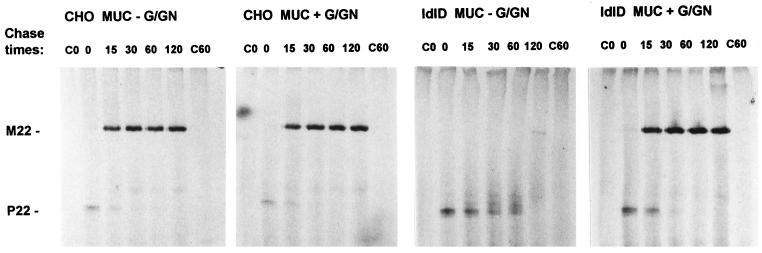
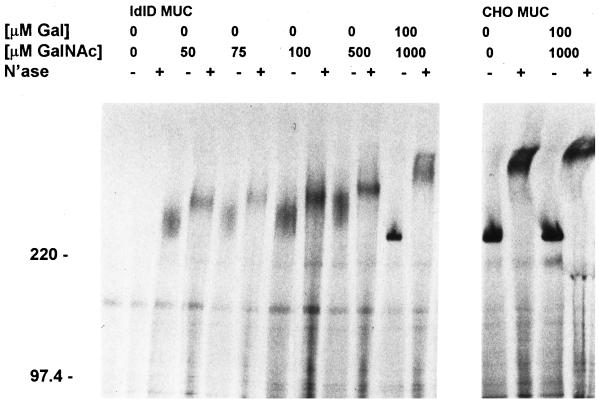
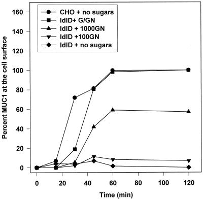
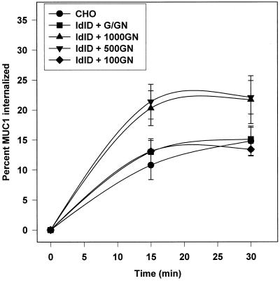
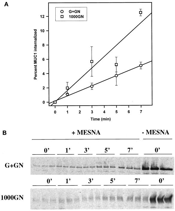
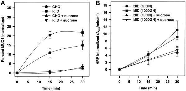
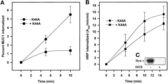
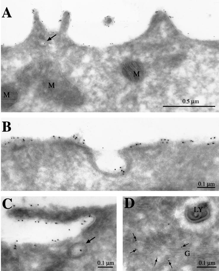
MUC1 is a mucin-like type 1 transmembrane protein associated with the apical surface of epithelial cells. In human tumors of epithelial origin MUC1 is overexpressed in an underglycosylated form with truncated O-glycans and accumulates in intracellular compartments. To understand the basis for this altered subcellular localization, we compared the synthesis and trafficking of various glycosylated forms of MUC1 in normal (Chinese hamster ovary) cells and glycosylation-defective (ldlD) cells that lack the epimerase to make UDP-Gal/GalNAc from UDP-Glc/GlcNAc. Although the MUC1 synthesized in ldlD cells was rapidly degraded, addition of GalNAc alone to the culture media resulted in stabilization and near normal surface expression of MUC1 with truncated but sialylated O-glycans. Interestingly, the initial rate of endocytosis of this underglycosylated MUC1 was stimulated by twofold compared with fully glycosylated MUC1. However, the half-lives of the two forms were not different, indicating that trafficking to lysosomes was not affected. Both the normal and stimulated internalization of MUC1 could be blocked by hypertonic media, a hallmark of clathrin-mediated endocytosis. MUC1 endocytosis was also blocked by expression of a dominant-negative mutant of dynamin-1 (K44A), and MUC1 was observed in both clathrin-coated pits and vesicles by immunoelectron microscopy of ultrathin cryosections. Our data suggest that the subcellular redistribution of MUC1 in tumor cells could be a direct result of altered endocytic trafficking induced by its aberrant glycosylation; potential models are discussed. These results also implicate a new role for O-glycans on mucin-like membrane proteins entering the endocytic pathway through clathrin-coated pits.


Figures

Figure 1
Figure 1
MUC1 maturation in ldlD cells is rescued by Gal and GalNAc. CHO and ldlD cells expressing human MUC1 were pulse labeled with [35S]Met/Cys for 15 min and chased for 0–120 min. Where indicated, both 100 μM Gal and 1000 μM GalNAc (+G/GN) were included in the starvation, pulse and chase media. MUC1 was immunoprecipitated from cell extracts and subjected to SDS-PAGE on a 3–10% polyacrylamide gradient gel before fluorography of the dried gel. C0 and C60 in each panel show immunoprecipitates from nontransfected control cells, which were labeled and then chased for 0 and 60 min, respectively. M22 (∼250 kDa) and P22 (130 kDa) denote the positions of the mature and propeptide forms of the recombinant MUC1 with 22 tandem repeats, respectively.
Figure 2
Figure 2
Truncated O-glycans on MUC1 can rescue its surface expression in ldlD cells. Both CHO and ldlD cells expressing MUC1 were pulsed with [35S]Met/Cys for 15 min and chased for 90 min. Varying levels of Gal and GalNAc were included in the starvation, pulse, and chase media as indicated. Immunoprecipitates of MUC1 from cell extracts were divided in half and incubated with or without neuraminidase. Numbers to the left of the gel correspond to the mobility of the molecular weight standards in kilodaltons. All samples were subjected to SDS-PAGE and fluorography.
Figure 3
Figure 3
MUC1 with truncated O-glycans reaches the cell surface with normal kinetics. Both CHO and ldlD cells expressing MUC1 were pulsed with [35S]Met/Cys for 15 min and chased for the times indicated before biotinylation of the cell surface. Varying levels of Gal and GalNAc were included in the starvation, pulse, and chase media as indicated (100 μM GalNAc, +100GN; 1000 μM GalNAc, +1000GN; 1000 μM GalNAc and 100 μM Gal, +G/GN). Biotinylated [35S]MUC1 was recovered with avidin-conjugated beads from the immunoprecipitates and subjected to SDS-PAGE for analysis of radioactive bands with a Bio-Rad phosphoimager system. Cell surface MUC1 levels in ldlD samples were normalized to the maximal level of cell surface [35S]MUC1 synthesized in ldlD cells in the presence of both Gal and GalNAc (+G/GN). The absolute levels of MUC1 expression in CHO and ldlD cells (+G/GN) are comparable (see Figure 5B).
Figure 4
Figure 4
MUC1 internalization from the cell surface is affected by its glycosylation state. CHO and ldlD cells expressing MUC1 were pulse labeled for 30 min and chased for 90 min before cell surface biotinylation on ice as described in MATERIALS AND METHODS. Varying levels of Gal (G, 100 μM) and GalNAc (GN, 100, 500, or 1000 μM) were included in the starvation, pulse and chase media as indicated. Cells were rapidly warmed to 37°C for the indicated times to allow internalization of surface MUC1 and rapidly cooled on ice, and remaining cell surface biotin was stripped with MESNA. Some samples were not treated with MESNA in order to determine the total amount of biotinylated MUC1 at t = 0. Internalized biotinylated MUC1 was recovered from the MUC1 immunoprecipitates with avidin-conjugated beads, and [35S]MUC1 was analyzed after SDS-PAGE using a phosphoimager. Data are plotted as the percent of total biotinylated [35S]MUC1 internalized at each time point and are presented as means ± SD of triplicate samples. Similar results were obtained in six experiments.
Figure 5
Figure 5
The initial rate of MUC1 endocytosis is enhanced by altered glycosylation. (A) The rate of endocytosis of [35S]MUC1 in ldlD cells, synthesized in the presence of either 1000 μM GalNAc (1000GN) with or without 100 μM Gal (G+GN), was characterized as described in MATERIALS AND METHODS and the legend to Figure 4. The mean of the percent endocytosis ± SEM for each time point for three experiments is shown. By paired Student's t test analysis, only the data at 7 min was significantly different (p = 0.015) between the two sugar conditions. Regression lines are drawn for each data set and indicate a 2.2-fold difference in the rate of endocytosis (p < 0.05). (B) A representative SDS-gel pattern for internalized [35S]MUC1 from a single experiment is shown, where n = 3 at each time point.
Figure 6
Figure 6
MUC1 internalization is inhibited by hypertonic media. (A) CHO and ldlD cells expressing MUC1 were pulse labeled for 30 min and chased for 90 min. GalNAc (1000 μM) was included in the starvation, pulse, and chase media for the ldlD cells. After biotinylation on ice, cells were rapidly warmed to 37°C in the presence of normal or hypertonic media (supplemented with 0.45 M sucrose) for the indicated times. The level of internalized MUC1 was determined as described in MATERIALS AND METHODS and the legend to Figure 4. (B) ldlD cells expressing MUC1 were preincubated for 2 h in media with 1000 μM GalNAc, with or without 100 μM Gal. Endocytosis of the fluid phase marker HRP, carried out in the presence (+ sucrose) or absence of hypertonic media, was determined as described in MATERIALS AND METHODS.
Figure 7
Figure 7
MUC1 internalization is dynamin dependent. (A) Clonal CHO cells expressing both MUC1 and hCAR were infected with adenoviruses expressing the tetracycline-repressible transactivating protein (AV-TA) and the dominant-negative mutant of dynamin-1 (+K44A, ▪). Controls (−K44A, ●) for the experiments were either cells infected with only AV-TA or cells infected with both viruses but incubated subsequently with the tetracyline analogue doxycycline (DOX) in the media. The following day, [35S]MUC1 endocytosis was determined as described in MATERIALS AND METHODS and the legend to Figure 5. The mean of the percent endocytosis ± SEM for each time point for six (t = 5 min) and seven (t = 10 min) experiments is shown. By paired Student's t test analysis, MUC1 endocytosis under the two conditions was significantly different at both time points (p ≤ 0.01). (B) CHO cells expressing MUC1 and infected as described above for (A) were grown overnight with (−K44A) or without (+K44A) DOX in the media before assay of HRP uptake as described in MATERIALS AND METHODS. Data are a representative experiment with the mean ± SD of triplicate samples at each time point. (C) Identical aliquots of adenoviral-infected cells from the experiment described in (B) were grown overnight with (+DOX) or without (−DOX) doxycycline and subjected to Western blot analysis with an anti-HA tag antibody to visualize HA-tagged dynamin-1 (K44A) under the two culture conditions.
Figure 8
Figure 8
Distribution of MUC1 in transfected CHO cells. Ultrathin cryosections of CHO cells expressing MUC1 were sequentially incubated with anti-MUC1 monclonal antibody VU-3-C6 and protein A conjugated to 5 nm colloidal gold. MUC1 was observed at the cell surface (A–C), in coated pits (B), and in coated invaginations and coated vesicles (marked with arrows in A and C, respectively). (D) MUC1 was also found in Golgi-associated vesicles. G, Golgi; LY, lysosome; M, mitochondria.
Figure 9
Figure 9
The endocytosis of pIgR in ldlD cells is unaffected by changes in glycosylation conditions or the presence of MUC1. The endocytosis of [35S]pIgR synthesized in the presence of either 1000 μM GalNAc, with (●) or without (▪) 100 μM Gal, in ldlD cells with (B) or without (A) MUC1 coexpression was characterized as described in MATERIALS AND METHODS. Endocytosis of dimeric 125I-labeled IgA prebound to pIgR in ldlD cell with (D) or without (C) coexpression of MUC1 was carried out after preincubation for 2 h with 1000 μM GalNAc, with (●) or without (▪) 100 μM Gal. Points are the mean of triplicate samples for representative experiments. Experiments were carried out 2–4 times with similar results. Error values at each point were <20% for both assays.

References

    1. Altschuler Y, Barbas SM, Terlecky LJ, Tang K, Hardy S, Mostov K, Schmid SL. Redundant and distinct functions for dynamin-1 and dynamin-2 isoforms. J Cell Biol. 1998;143:1871–1881. - PMC - PubMed
    1. Altschuler Y, Poland PA, Kinlough CL, Hughey RP. Role of endocytosis in the maturation of the membrane-associated mucin MUC1. Mol Biol Cell. 1997;8:301a.
    1. Barylko B, Binns D, Lin K-M, Atkinson MAL, Jameson DM, Yin HL, Albanesi JP. Synergistic activation of dynamin GTPase by Grb2 and phosphoinositides. J Biol Chem. 1998;273:3791–3797. - PubMed
    1. Bergelson JM, Cunningham JA, Droguett G, Kurt-Jones EA, Krithivas A, Hong JS, Horwitz MS, Crowell RL, Finberg RW. Isolation of a common receptor for coxsackie B viruses and adenoviruses 2 and 5. Science. 1997;275:1320–1323. - PubMed
    1. Bierhuizen MFA, Maemura K, Fukuda M. Expression of a differentiation antigen and poly-N-acetyllactosaminyl O-glycans directed by a cloned core 2 β-1,6-N-acetylglucosaminyltransferase. J Biol Chem. 1994;269:4473–4479. - PubMed

Publication types