. 2000 Mar;11(3):915-27.
doi: 10.1091/mbc.11.3.915.

Degradation of the transcription factor Gcn4 requires the kinase Pho85 and the SCF(CDC4) ubiquitin-ligase complex

Affiliations
Free PMC article

Degradation of the transcription factor Gcn4 requires the kinase Pho85 and the SCF(CDC4) ubiquitin-ligase complex

A Meimoun et al. Mol Biol Cell. 2000 Mar.
Free PMC article

Abstract

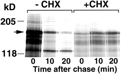
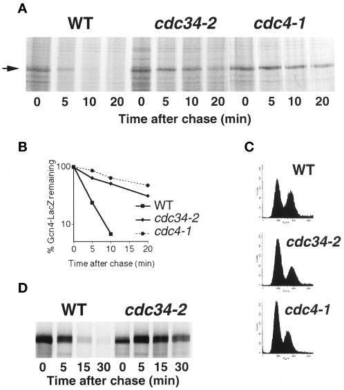
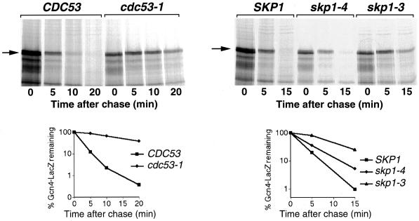
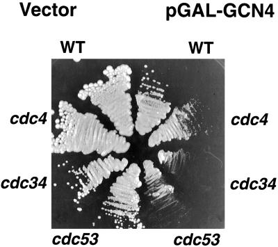
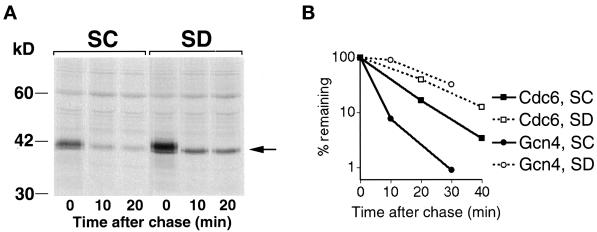
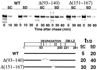
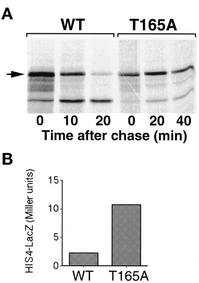
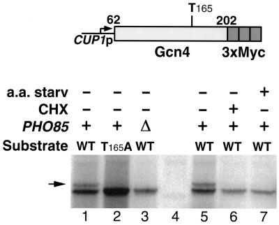
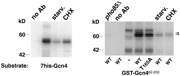
Gcn4, a yeast transcriptional activator that promotes the expression of amino acid and purine biosynthesis genes, is rapidly degraded in rich medium. Here we report that SCF(CDC4), a recently characterized protein complex that acts in conjunction with the ubiquitin-conjugating enzyme Cdc34 to degrade cell cycle regulators, is also necessary for the degradation of the transcription factor Gcn4. Degradation of Gcn4 occurs throughout the cell cycle, whereas degradation of the known cell cycle substrates of Cdc34/SCF(CDC4) is cell cycle regulated. Gcn4 ubiquitination and degradation are regulated by starvation for amino acids, whereas the degradation of the cell cycle substrates of Cdc34/SCF(CDC4) is unaffected by starvation. We further show that unlike the cell cycle substrates of Cdc34/SCF(CDC4), which require phosphorylation by the kinase Cdc28, Gcn4 degradation requires the kinase Pho85. We identify the critical target site of Pho85 on Gcn4; a mutation of this site stabilizes the protein. A specific Pho85-Pcl complex that is able to phosphorylate Gcn4 on that site is inactive under conditions under which Gcn4 is stable. Thus, Cdc34/SCF(CDC4) activity is constitutive, and regulation of the stability of its various substrates occurs at the level of their phosphorylation.


Figures

Figure 1
Figure 1
Inhibition of translation stabilizes Gcn4. Gcn4–LacZ was expressed from the CUP1 promoter of plasmid KB449. +CHX: cells were preincubated for 30 min in 0.5 μg/ml cycloheximide and then labeled and chased in the presence of the drug. The arrow on the left indicates Gcn4–LacZ.
Figure 2
Figure 2
Gcn4 degradation is inhibited in cdc34 and cdc4 mutants. (A) Pulse-and-chase analysis of Gcn4–LacZ expressed from the CUP1 promoter of plasmid KB449 in strains KY204, KY205, and KY301. The cultures were shifted from 30 to 37°C, 20 min before the labeling. (B) Quantitation of the experiment shown in (A). (C) Fluorescence-activated cell sorter analysis of cells at the time of labeling. (D) Pulse–chase analysis of Gcn4–-LacZ expressed from the GAL1 promoter of plasmid KB64 in α-factor–arrested cells. Cells were arrested with α-factor at the permissive temperature and then shifted to 37° for 30 min before labeling.
Figure 3
Figure 3
Gcn4 degradation is inhibited in cdc53 and skp1 mutants. Pulse–chase analysis of Gcn4–-LacZ expressed from the CUP1 promoter of plasmid KB449 in strains W303 and MTY740 (left) or YPH1015, YPH1161, and YPH1172 (right). The cultures were shifted from 30 to 37°C, 30 min before labeling.
Figure 4
Figure 4
Effect of Gcn4 overexpression on the growth of various SCF mutants. Plasmid KB496, expressing Gcn4 under the strong GAL1–CYC1 hybrid promoter, or the vector plasmid pLGSD5, were transformed into the indicated strains. The strains were streaked on selective galactose plates and incubated for 3 d at 30°C.
Figure 5
Figure 5
Starvation does not affect degradation of two other SCFCDC4 substrates. (A) Degradation of Sic1. Cells carrying plasmids KB733 (CUP1-SIC1) and pGAL1-CLN2(4T3S)HA (Lanker et al., 1996) were arrested in S-phase with hydroxyurea, induced with galactose to produce Cln2, and subjected to pulse–chase analysis without (SC) or with (SD) prior amino acid starvation. The Sic1 protein was visualized by immunoprecipitation with a specific antibody. The arrow on the right indicates Sic1. (B) Degradation of Cdc6–LacZ versus Gcn4–LacZ. Cells carrying plasmid KB448 (pGAL1–CDC6–LacZ) or KB64 (pGAL1–GCN4–LacZ) were grown in galactose, arrested in G1 with α-factor, and subjected to pulse–chase analysis without (SC) or with (SD) prior amino acid starvation (30 min in medium lacking amino acids). The lacZ fusion proteins were visualized by immunoprecipitation with a specific LacZ antibody. The graph depicts the quantitation of the fusion protein band by phosphorimager.
Figure 6
Figure 6
Effect of deletions in Gcn4 on the sensitivity to starvation. Full-length Gcn4 and two deletions fused to LacZ were subjected to pulse–chase analysis without (SC) or with (SD) prior mild starvation (15 min in medium lacking amino acids). Note the difference in time scale. t½ indicates the half-lives of the three proteins under both conditions as derived from quantitation of the data by phosphorimager. “DEGRADATION” denotes the minimal region of Gcn4 required for degradation, and “I” and “II” indicate the two domains of that region, as defined by deletion analysis (see text). “DB-LZ” denotes the DNA binding domain and the leucine zipper domain of Gcn4.
Figure 7
Figure 7
Degradation of Gcn4 in pho85Δ cells. (A) Pulse–chase analysis of Gcn4–LacZ expressed from the CUP1 promoter of plasmid KB449 in W303 and DY4535 cells. (B) Quantitation of the experiment shown in (A). (C) Pulse–chase analysis of native, endogenous Gcn4 in W303 and DY4535 cells. The lane marked “C” indicates an extract from a gcn4Δ strain. The anomalous migration of native Gcn4 (shown here) as well as of recombinant Gcn4 (Figures 10 and 11), a 33-kDa protein, has been noted before (Hope and Struhl, 1985). Note that a doublet of cross-reacting bands migrates slightly slower than Gcn4. (D) Expression of His4–LacZ from a HIS4 promoter derivative that is exclusively dependent on Gcn4 for activity (deletion number 203; Nagawa and Fink, 1985) in W303 and DY4535 cells. Overnight cultures were diluted and grown for 6 h to early log phase, and the β-galactosidase activity of the cultures was measured as described (Daignan-Fornier and Fink, 1992). (E) Degradation of the T105A mutant of Gcn4 in W303 and DY4535 cells. Gcn4(T105A)-LacZ was expressed from the GAL promoter of plasmid KB358 (Kornitzer et al., 1994).
Figure 8
Figure 8
A mutation at residue Thr165 stabilizes Gcn4. (A) Pulse–chase analysis of wild-type (KB149) and mutant (KB854) Gcn4–LacZ expressed from the GAL promoter in W303 cells. Note the difference in time scale. (B) Expression of His4–LacZ from the full-length HIS4 promoter (plasmid pFN6; Nagawa and Fink, 1985). The strain used, bas1-2 bas2-2 gcn4Δ1 (KY26), is defective in both HIS4 transcription factors (Arndt et al., 1987). His4–LacZ expression is exclusively dependent on the plasmid-borne GCN4 alleles expressed from the ADE8 promoter of plasmids KB105 (WT) or KB853 (T165A). The β-galactosidase assay was performed as for Figure 7D.
Figure 9
Figure 9
In vivo phosphorylation of a fragment of Gcn4 encompassing residues 62–202 fused to a triple Myc epitope and expressed from the CUP1 promoter. The upper phosphorylation band (indicated by an arrow) is 30–40% as intense as the lower band in lanes 1 and 5 and is undetectable in the other lanes, even upon prolonged exposure (we estimate the limit of detection at <2% of the main band). Lane 4 is a no-tag control. CHX, cycloheximide treatment (1 μg/ml, 30 min); a.a. starv, 30-min incubation in YNB medium lacking all amino acids.
Figure 10
Figure 10
In vitro phosphorylation of Gcn4 by Pho85-Pcl1. Different substrates (indicated at the bottom) were incubated with wild-type or catalytically inactive (E53A) GST-Pho85, with or without GST-Pcl1. Asterisk indicates background phosphorylation on bacterial proteins copurifying with the recombinant kinase and cyclin. The two arrowheads indicate the two phosphorylation bands of GST-Gcn462–202. The two weak signals (<10% intensity) visible over the main band in the T165A mutant presumably represent phosphorylation at alternative sites. The right panel shows that GST alone is not phosphorylated by Pho85-Pcl1 (the GST protein band would be expected to migrate slightly faster than the 30-kDa marker).
Figure 11
Figure 11
In vitro phosphorylation of Gcn4 by ha-Pcl1–associated kinase activity. Different substrates (indicated at the bottom) were incubated with Protein A-agarose beads carrying Pcl1-associated kinase activity immunoprecipitated from various cell extracts. pho85Δ indicates that the extract was made from a pho85 mutant strain; all other extracts were from wild-type cells. starv. and CHX refer to the same treatments as in Figure 9. no Ab refers to a blank immunoprecipitation in the absence of antibody. The two arrowheads at the right indicate the two phosphorylation bands of the GST-Gcn462–202 substrate.
Figure 12
Figure 12
A model of the proposed pathway of regulation of Gcn4 degradation by starvation. See text for details.

References

    1. Alani E, Cao L, Kleckner N. A method for gene disruption that allows repeated use of URA3 selection in the construction of multiply disrupted yeast strains. Genetics. 1987;116:541–545. - PMC - PubMed
    1. Andrews B, Measday V. The cyclin family of budding yeast: abundant use of a good idea. Trends Genet. 1998;14:66–72. - PubMed
    1. Arndt KT, Styles CA, Fink GR. Multiple global regulators control HIS4 transcription in yeast. Science. 1987;237:874–880. - PubMed
    1. Ausubel FM, Brent R, Kingston RE, Moore DD, Seidman JG, Smith JA, Struhl K. Current Protocols in Molecular Biology. New York: John Wiley & Sons; 1989.
    1. Bai C, Sen P, Hoffman K, Ma L, Goebl M, Harper JW, Elledge SJ. SKP1 connects cell cycle regulators to the ubiquitin proteolysis machinery through a novel motif, the F-box. Cell. 1996;86:263–274. - PubMed

Publication types

MeSH terms