. 2000 Mar;11(3):1061-76.
doi: 10.1091/mbc.11.3.1061.

Transforming growth factor beta(1) selectively inhibits the cyclic AMP-dependent proliferation of primary thyroid epithelial cells by preventing the association of cyclin D3-cdk4 with nuclear p27(kip1)

Affiliations
Free PMC article

Transforming growth factor beta(1) selectively inhibits the cyclic AMP-dependent proliferation of primary thyroid epithelial cells by preventing the association of cyclin D3-cdk4 with nuclear p27(kip1)

F Depoortere et al. Mol Biol Cell. 2000 Mar.
Free PMC article

Abstract

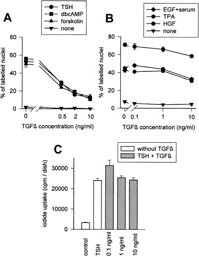
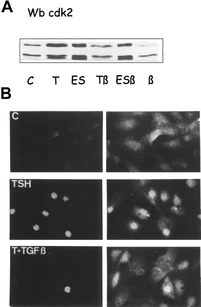
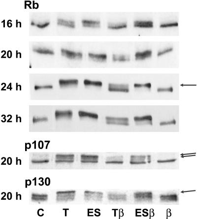
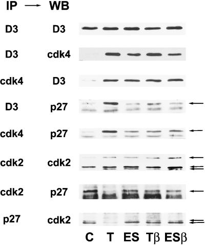
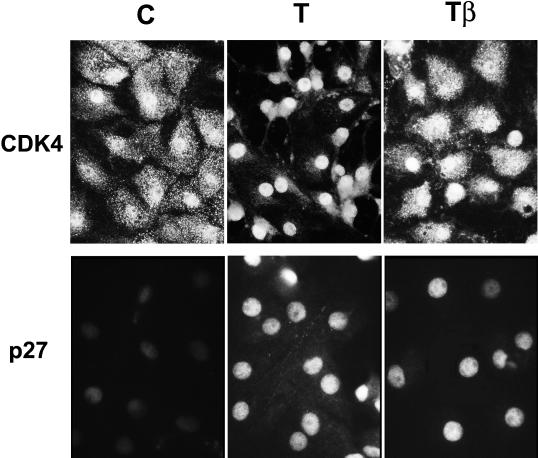
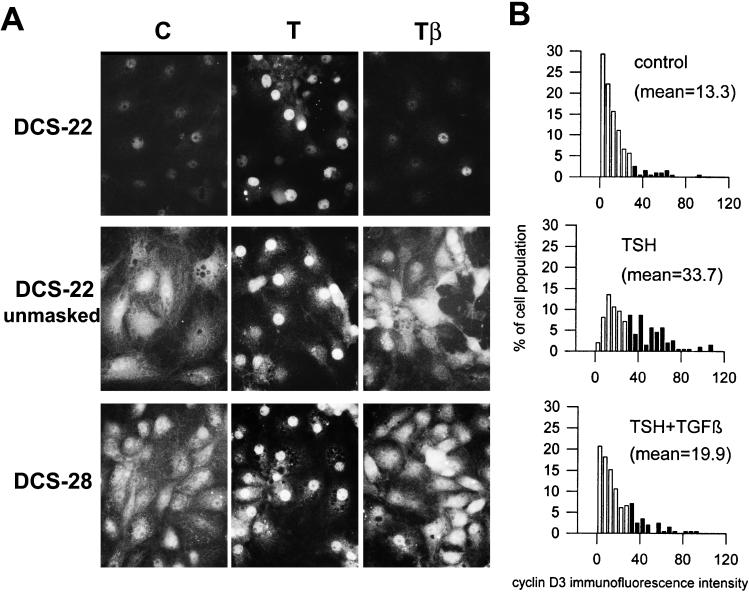
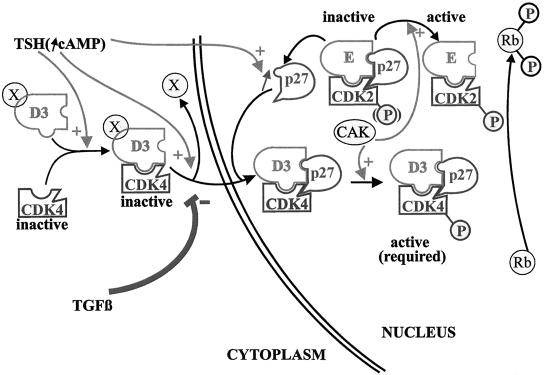
Dog thyroid epithelial cells in primary culture constitute a physiologically relevant model of positive control of DNA synthesis initiation and G0-S prereplicative phase progression by cAMP as a second messenger for thyrotropin (thyroid-stimulating hormone [TSH]). As previously shown in this system, the cAMP-dependent mitogenic pathway differs from growth factor cascades as it stimulates the accumulation of p27(kip1) but not cyclins D. Nevertheless, TSH induces the nuclear translocations and assembly of cyclin D3 and cdk4, which are essential in cAMP-dependent mitogenesis. Here we demonstrate that transforming growth factor beta(1) (TGFbeta(1)) selectively inhibits the cAMP-dependent cell cycle in mid-G1 and various cell cycle regulatory events, but it weakly affects the stimulation of DNA synthesis by epidermal growth factor (EGF), hepatocyte growth factor, serum, and phorbol esters. EGF+serum and TSH did not interfere importantly with TGFbeta receptor signaling, because they did not affect the TGFbeta-induced nuclear translocation of Smad 2 and 3. TGFbeta inhibited the phosphorylation of Rb, p107, and p130 induced by TSH, but it weakly affected the phosphorylation state of Rb-related proteins in EGF+serum-treated cells. TGFbeta did not inhibit c-myc expression. In TSH-stimulated cells, TGFbeta did not affect the expression of cyclin D3, cdk4, and p27(kip1), nor the induced formation of cyclin D3-cdk4 complexes, but it prevented the TSH-induced relocalization of p27(kip1) from cdk2 to cyclin D3-cdk4. It prevented the nuclear translocations of cdk4 and cyclin D3 without altering the assembly of cyclin D3-cdk4 complexes probably formed in the cytoplasm, where they were prevented from sequestering nuclear p27(kip1) away from cdk2. This study dissociates the assembly of cyclin D3-cdk4 complexes from their nuclear localization and association with p27(kip1). It provides a new mechanism of regulation of proliferation by TGFbeta, which points out the subcellular location of cyclin D-cdk4 complexes as a crucial factor integrating mitogenic and antimitogenic regulations in an epithelial cell in primary culture.


Figures

Figure 1
Figure 1
Selective inhibition by TGFβ of the cAMP-dependent stimulation of DNA synthesis. Quiescent 4-day-old dog thyrocytes were stimulated for 48 h with either (A) TSH (1 mU/ml), forskolin (10−5 M), or dibutyryl cAMP (10−4 M), or (B) EGF (25 ng/ml), HGF (40 ng/ml), or TPA (10 ng/ml), or none, in combination or not with different TGFβ1 concentrations. BrdU was present for the last 24 h, and the percentage of BrdU-labeled cells was determined. (C) Iodide uptake was measured in parallel. Four-day-old dog thyrocytes were stimulated for 48 h by TSH (1 mU/ml) with or without various TGFβ1 concentrations or remained in control condition. Cells were then incubated for 2 h with 131I-labeled Na (10−6 M, 2 μCi/ml) and mercaptomethylimidazol (1 mM), and the cell radioactivity was measured as described (Taton et al., 1993).
Figure 2
Figure 2
Dog thyrocytes are sensitive to TGFβ1 inhibition during G1. (●) Quiescent 4-day-old dog thyrocytes were stimulated at 0 h with TSH (1 mU/ml) in the presence of BrdU and the cumulative BrdU-labeling index was determined at the indicated times. (○) At the indicated times after TSH administration at 0 h in the presence of BrdU, TGFβ1 (2 ng/ml) was added and the incorporation of BrdU was allowed to proceed until 48 h after TSH addition, at which time all cultures were fixed, and the BrdU-labeling index was determined. The 48-h labeling index without TGFβ was 54% (normalized to 100% on the axis; ●) and the maximal inhibition by TGFβ1 (normalized to 100% on the axis; ○) was 70% inhibition of the nuclear labeling.
Figure 3
Figure 3
Inhibition by TGFβ of TSH-induced cell cycle progression (A) and accumulation of cell cycle regulatory proteins (B). Four-day-old dog thyrocytes were stimulated at 0 h with TSH (1 mU/ml) or remained in control (C) condition. TGFβ1 (2 ng/ml) (β) was administrated at the same time or 15 h after TSH. (A) Kinetics of cell cycle progression evaluated by the determination of the percentage of PCNA-positive cells identified as described (Baptist et al., 1993) in G1 and S phases. PCNA-negative cells are either in G0 or at a G1 stage before PCNA appearance. (B) Accumulation of cdk4, cdk2, cyclin A, and cdc2 assessed by Western blotting in the same experiment.
Figure 4
Figure 4
Inhibition by TGFβ of the TSH-stimulated phosphorylation (A) and nuclear translocation (B) of cdk2. Four-day-old dog thyrocytes were stimulated by TSH (1 mU/ml) or EGF (25 ng/ml) + serum (10%) (ES) alone or in combination with TGFβ1 (2 ng/ml) (β). (A) Activating Thr160 phosphorylation of cdk2 reflected by its downward electrophoretic shift (Gu et al., 1992) demonstrated by Western blotting from cells stimulated for 32 h. (B) Double immunofluorescence labeling of PCNA used as a cell cycle marker (left panels) and cdk2 (right panels) 26 h after cell stimulation. Notice the increased nuclear labeling of cdk2 in PCNA-positive cells observed in many TSH-stimulated cells but few cells treated with TSH+TGFβ.
Figure 5
Figure 5
Nuclear translocation of Smad 2 and 3 induced by TGFβ. Four-day-old dog thyrocytes were stimulated for 20 h with TSH (1 mU/ml) (T), EGF (25 ng/ml) + serum (10%) (ES) or remained in control (C) condition. They were then incubated for 2 h with TGFβ1 (10 ng/ml) before fixation and immunofluorescent labeling using an antibody against Smad 2 and 3 proteins.
Figure 6
Figure 6
Accumulation of c-myc mRNA in dog thyrocytes analyzed by Northern blotting. Quiescent 4-day-old cells were stimulated for 1 or 3 h with TSH (T), EGF+serum (ES) with or without TGFβ or by TGFβ (β) alone or remained in control (C) condition. Northern blots were prepared with 10 μg of glyoxal denatured total RNA. Acridine orange was performed to assess that equal amount of RNA were loaded in independent lanes.
Figure 7
Figure 7
TGFβ inhibits the TSH-stimulated phosphorylation of proteins of the Rb family, as detected by their electrophoretic shifts evidenced by Western blotting. Quiescent 4-day-old dog thyrocytes were stimulated during various times with TSH (T) (1 mU/ml), EGF (25 ng/ml) + serum (ES) with or without TGFβ, or by TGFβ alone (2 ng/ml) (β) or remained in control (C) condition. The modulated bands corresponding to the slow-migrating, hyperphosphorylated forms of Rb, p107, and p130 are indicated by arrows.
Figure 8
Figure 8
Kinetics of accumulation of cyclin D3 and p27kip1 detected by Western blotting. Quiescent 4-day-old dog thyroid cells were stimulated for the indicated times using TSH (1 mU/ml) (T) with or without TGFβ (2 ng/ml) (β), EGF (25 ng/ml) + serum (ES) or remained in control (C) condition.
Figure 9
Figure 9
TGFβ inhibits the TSH-induced relocalization of p27kip1 from cdk2 to cyclin D3–cdk4, but not the assembly of cyclin D3–cdk4 complexes. Quiescent dog thyrocytes were stimulated for 20 h with TSH (1 mU/ml) (T) or EGF (25 ng/ml) + serum (10%) (ES) with or without TGFβ (2 ng/ml) (β) or remained in control (C) condition. Immunoprecipitations (IP) were carried out from equal amounts of cell extracts with antibodies against cyclin D3, cdk4, cdk2, and p27kip1, followed by separation by SDS-PAGE. The proteins were then transferred to nitrocellulose membranes, followed by Western blotting (WB) with the indicated antibodies. The position of p27kip1 and the different forms of cdk2 are indicated by arrows in the respective panels.
Figure 10
Figure 10
TGFβ inhibits the TSH-induced nuclear translocation of cdk4 but does not affect the nuclear location of p27kip1. Quiescent 4-day-old dog thyrocytes were stimulated for 20 h with TSH (1 mU/ml) (T) with or without TGFβ (2 ng/ml) (β) or remained in control (C) condition. Cells were then processed for cdk4 or p27kip1 immunofluorescent staining. Labeled cells were photographed using a 50× immersion lens (cdk4) or a 100× lens (p27).
Figure 11
Figure 11
TGFβ inhibits the epitope unmasking and nuclear translocation of cyclin D3 induced by TSH. Quiescent 4-day-old dog thyrocytes were stimulated for 20 h with TSH (1 mU/ml) (T) with or without TGFβ (2 ng/ml) (β) or remained in control (C) condition. Cells were then processed for cyclin D3 immunofluorescent staining using the DCS-22 or DCS-28 monoclonal antibodies. For cyclin D3 detection using DCS-22, an epitope unmasking treatment by a mild trypsin digestion of fixed cells was applied (unmasked) or not. (A) Labeled cells were photographed using a 50× immersion lens. (B) Distribution of nuclear cyclin D3 immunofluorescence intensities within the cell population as measured by photometry. Cyclin D3 was detected using DCS-22 without the application of the trypsin unmasking treatment. Values of nuclear immunofluorescences exceeding the weak to moderate ones recorded in 90% of unstimulated control cells were scored positive and are shown as filled bars. Average values of immunofluorescence intensity are indicated for each treatment.
Figure 12
Figure 12
Double immunofluorescent labeling of cyclin D3 (using DCS-22 without unmasking treatment) or cdk4, and PCNA used as a cell cycle marker. Quiescent dog thyrocytes were stimulated for 26 h with TSH (1 mU/ml) + TGFβ (2 ng/ml). Large arrows and medium size arrows show cells identified, respectively, in late G1 and S phases as described previously (Baptist et al., 1993) (the speckled appearance of PCNA-labeled S-phase nuclei was clearly seen at microscope but not in these digitalized micrographs). Small arrows show G0/G1 cells with a low PCNA staining. The microscopical fields were selected to contain cells that escape the TGFβ inhibition of proliferation. Notice that all the PCNA-positive cycling cells display intense nuclear stainings of cyclin D3 and cdk4.
Figure 13
Figure 13
The subcellular localization of cyclin D3 and cdk4 integrates the antagonistic cell cycle effects of TSH and TGFβ. TSH does not stimulate cyclin D3 accumulation, but it assembles cyclin D3–cdk4 complexes, enhances the levels of p27kip1, and induces the nuclear translocation of cyclin D3–cdk4, which correlates with the exposition of a cyclin D3 epitope (depicted by the displacement of the X element). Cyclin D3–cdk4 complexes are stabilized in the nucleus by their binding to p27kip1, which thus might serve as a nuclear anchor. This nuclear translocation of cdk4 is assumed to be required for its phosphorylation by nuclear CAK and for access to Rb. TGFβ does not inhibit the assembly of cyclin D3–cdk4 complexes, nor p27kip1 accumulation, but it prevents the epitope unmasking of cyclin D3 and the nuclear translocation of cyclin D3–cdk4. Consequently these complexes are prevented from encountering p27kip1 and thus sequestering it away from the initial pool of nuclear cdk2 complexes. Cyclin D3–cdk4 is thus maintained in an inactive state by its cytoplasmic location, and nuclear cdk2 is inhibited by its association with p27kip1. The further import of cdk2 from the cytoplasm induced by TSH but repressed by TGFβ as a likely indirect consequence of this mechanism, is not depicted.

References

    1. Alexandrow MG, Moses HL. Transforming growth factor beta 1 inhibits mouse keratinocytes late in G1 independent of effects on gene transcription. Cancer Res. 1995;55:3928–3932. - PubMed
    1. Baptist M, Dumont JE, Roger PP. Demonstration of cell cycle kinetics in thyroid primary culture by immunostaining of proliferating cell nuclear antigen: differences in cyclic AMP-dependent and -independent mitogenic stimulations. J Cell Sci. 1993;105:69–80. - PubMed
    1. Baptist M, Dumont JE, Roger PP. Intercellular heterogeneity of early mitogenic events: cAMP generalizes the EGF effect on c-Fos protein appearance but not on MAP kinase phosphorylation and nuclear translocation in dog thyroid epithelial cells. Exp Cell Res. 1995;221:160–171. - PubMed
    1. Baptist M, Lamy F, Gannon J, Hunt T, Dumont JE, Roger PP. Expression and subcellular localization of CDK2 and cdc2 kinases and their common partner cyclin A in thyroid epithelial cells: comparison of cyclic AMP-dependent and -independent cell cycles. J Cell Physiol. 1996;166:256–273. - PubMed
    1. Barnard JA, Lyons RM, Moses HL. The cell biology of transforming growth factor beta. Biochim Biophys Acta. 1990;1032:79–87. - PubMed

Publication types

MeSH terms

Substances