

Crystal structures of ligand complexes of P450eryF exhibiting homotropic cooperativity
- PMID: 10716705
- PMCID: PMC16190
- DOI: 10.1073/pnas.97.7.3050
Crystal structures of ligand complexes of P450eryF exhibiting homotropic cooperativity
Abstract
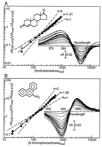
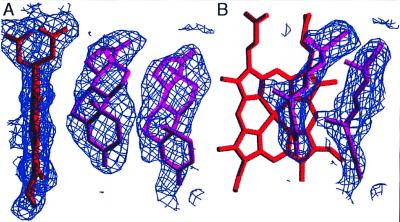
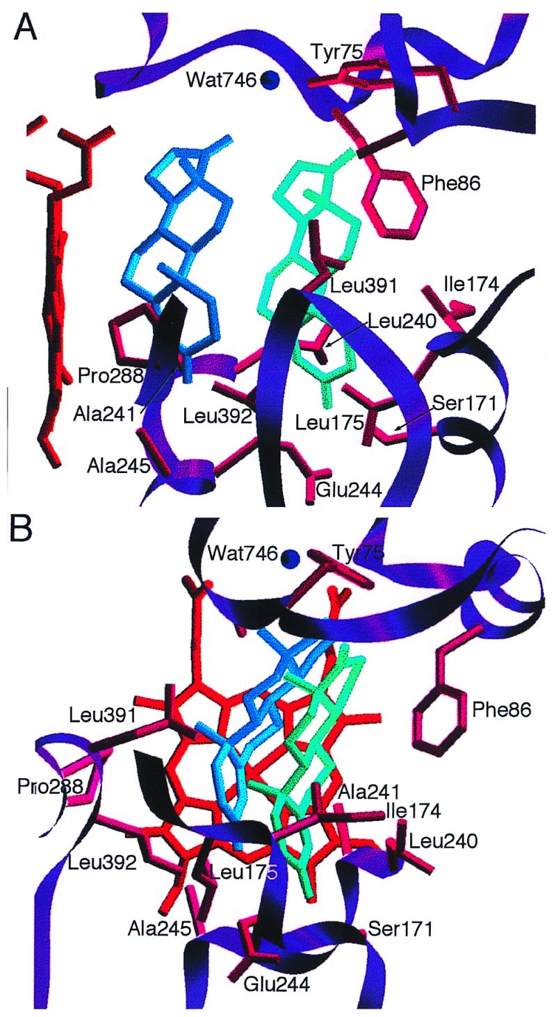
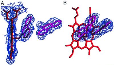
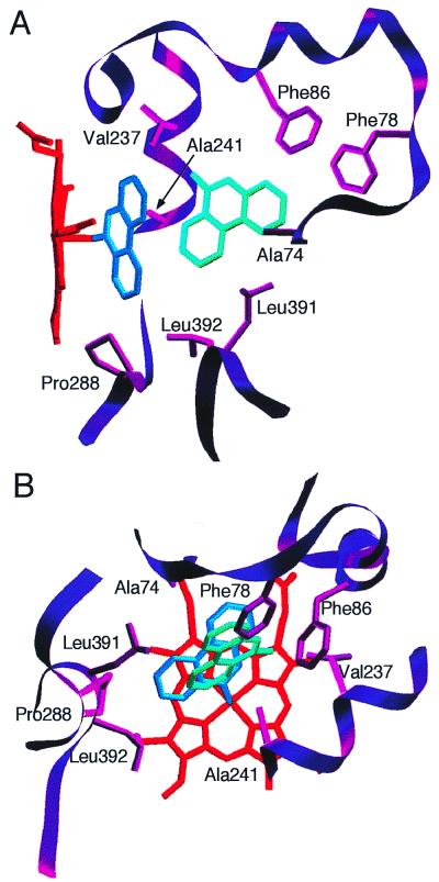
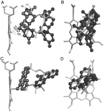
Several mammalian cytochrome P450 (P450) isoforms demonstrate homotropic cooperativity with a number of substrates, including steroids and polycyclic aromatic hydrocarbons. To identify structural factors contributing to steroid and polycyclic aromatic hydrocarbon binding to P450 enzymes and to determine the location of the allosteric site, we investigated interactions of the macrolide hydroxylase P450eryF from Saccharopolyspora erythraea with androstenedione and 9-aminophenanthrene. Spectroscopic binding assays indicate that P450eryF binds androstenedione with an affinity of 365 microM and a Hill coefficient of 1.31 +/- 0.6 and coordinates with 9-aminophenanthrene with an affinity of 91 microM and a Hill coefficient of 1.38 +/- 0.2. Crystals of complexes of androstenedione and 9-aminophenanthrene with P450eryF were grown and diffracted to 2.1 A and 2.35 A, respectively. Electron density maps indicate that for both complexes two ligand molecules are simultaneously present in the active site. The P450eryF/androstenedione model was refined to an r = 18.9%, and the two androstenedione molecules have similar conformations. The proximal androstenedione is positioned such that the alpha-face of carbon-6 is closest to the heme iron, and the second steroid molecule is positioned 5.5 A distal in the active site. The P450eryF/9-aminophenanthrene model was refined to an r = 19.7% with the proximal 9-aminophenanthrene coordinated with the heme iron through the 9-amino group and the second ligand positioned approximately 6 A distal in the active site. These results establish that homotropic cooperativity in ligand binding can result from binding of two substrate molecules within the active site pocket without major conformational changes in the protein.
Figures
References
-
- Ortiz de Montellano P R, editor. Cytochrome P450: Structure, Mechanism, and Biochemistry. New York: Plenum; 1986.
-
- Gunsalus I C, Pederson T C, Sligar S G. Annu Rev Biochem. 1975;44:377–407. - PubMed
-
- Hayaishi O. Molecular Mechanisms of O2 Activation. New York: Academic; 1974.
-
- Porter T D, Coon M J. FASEB J. 1992;6:669–673. - PubMed
-
- Guengerich F P. FASEB J. 1992;6:745–748. - PubMed
Publication types
MeSH terms
Substances
Associated data
- Actions
- Actions
LinkOut - more resources
Full Text Sources
Molecular Biology Databases
