. 2000 Mar 28;97(7):3336-41.
doi: 10.1073/pnas.97.7.3336.

Abnormal development of Purkinje cells and lymphocytes in Atm mutant mice

Affiliations

Abnormal development of Purkinje cells and lymphocytes in Atm mutant mice

P R Borghesani et al. Proc Natl Acad Sci U S A. .

Abstract

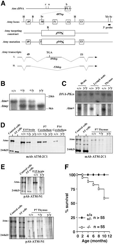
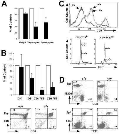
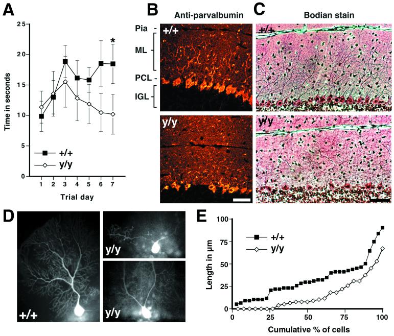
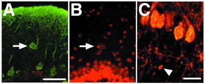
Motor incoordination, immune deficiencies, and an increased risk of cancer are the characteristic features of the hereditary disease ataxia-telangiectasia (A-T), which is caused by mutations in the ATM gene. Through gene targeting, we have generated a line of Atm mutant mice, Atm(y/y) mice. In contrast to other Atm mutant mice, Atm(y/y) mice show a lower incidence of thymic lymphoma and survive beyond a few months of age. Atm(y/y) mice exhibit deficits in motor learning indicative of cerebellar dysfunction. Even though we found no gross cerebellar degeneration in older Atm(y/y) animals, ectopic and abnormally differentiated Purkinje cells were apparent in mutant mice of all ages. These findings establish that some neuropathological abnormalities seen in A-T patients also are present in Atm mutant mice. In addition, we report a previously unrecognized effect of Atm deficiency on development or maintenance of CD4(+)8(+) thymocytes. We discuss these findings in the context of the hypothesis that abnormal development of Purkinje cells and lymphocytes contributes to the pathogenesis of A-T.


Figures

Figure 1
Figure 1
Generation of Atmy/y mice and Atm expression. (A) Location of the Atmy mutation and other reported murine Atm mutations; a (14), b (15, 16), and c (13). B, BamHI; H, HindIII; Bg, BglII; Sc, SacI; Xb, XbaI; and Neor, neomycin-resistance gene. (B) Southern blot analysis of Atmy mutation. BamHI-digested DNA is probed with the indicated 3′ probe (A). (C) Northern blot analysis of Atm expression. Total RNA from brain, lymph node, and thymus of wild-type (+/+) and Atmy/y (y/y) mice were probed with an Atm cDNA fragment (Lower) and reprobed with a DNA-PKcs cDNA probe (Upper) to confirm equal RNA loading. (D and E) Western blot analysis of Atm expression in Atmy/y tissues using antibodies raised against the C terminus (D) and N terminus (E) of the ATM protein. The bands at ≈200 kDa and ≈300 kDa are likely nonspecific since they are present in human and murine samples of both genotypes. (F) Kaplan–Meier survival curve of Atmy/y mice (n = 55).
Figure 2
Figure 2
Abnormal T lymphocyte development in Atmy/y mice. (A) Average pairwise comparisons of weight, total numbers of thymocytes, and splenocytes from 10 same-sex littermates pairs 1–5 mo old. (B) (Upper) The average reduction of the total number of each Atmy/y thymocyte subpopulations (DN, CD48; DP; CD4+8+; CD4+SP, CD4+8; CD8+SP, CD48+) from seven littermate pairs. (Lower) FACS analysis of T cell development in Atmy/y thymuses. (C) Histogram of CD3 surface expression in Atmy/y thymocytes (y/y). (Lower) FSC (forward site scatter) using a lymphocyte gate on CD3low and CD3int DP cells, respectively. (D) Splenic T cells in Atmy/y mice (y/y) were reduced in numbers, but their surface phenotype (TCRβ/CD3 expression) appeared to be normal. Numbers indicate the percentage of splenic T cells (CD3+) and B cells (B220+).
Figure 3
Figure 3
Motor-learning deficits and dendritic changes in Atmy/y mice. (A) Performance of wild-type mice but not Atmy/y mice on an accelerating rotating rod improved over the trial period (P < 0.01 by repeated-measure ANOVA, regression slope = 1.21 s/day, P < 0.05), and by the seventh trial day, wild-type mice performed better than Atmy/y mice (P < 0.02, Mann–Whitney rank sum test). (B) Parvalbumin immunohistochemistry of midsagittal cerebellar sections from 5-mo-old wild-type (Upper) and Atmy/y mice (Lower) reveals the altered dendritic arborization of Purkinje cells in Atmy/y mice. (C) This can also be observed in Bodian-stained sections from 1-yr-old mice, wild-type (Upper), and Atmy/y (Lower). (D) Lucifer yellow-injected wild-type and Atmy/y Purkinje cells displaying either the prototypic morphology (+/+) or premature dendritic branching (y/y), which can result in multiple dendrites emanating from the soma (upper y/y cell). (E) Cumulative percent graph of stem dendrite length; 7 of 26 Atmy/y Purkinje cells have multiple dendrites. (Scale bar in B and C is 50 μm.)
Figure 4
Figure 4
(A and B) An ectopic Purkinje cell in an Atmy/y mouse. Ectopic cells were located and counted by double labeling for calbindin immunoreactivity (A) and propidium iodide staining (B). (C) Varicosity along Purkinje cell axon. (Scale bar in A is 50 μm and in C is 20 μm.)

References

    1. Gatti R A, Boder E, Vinters H V, Sparkes R S, Norman A, Lange K. Medicine. 1991;70:99–117. - PubMed
    1. Boder E. Kroc Found Ser. 1985;19:1–63. - PubMed
    1. Savitsky K, Bar-Shira A, Gilad S, Rotman G, Ziv Y, Vanagaite L, Tagle D A, Smith S, Uziel T, Sfez S, et al. Science. 1995;268:1749–1753. - PubMed
    1. Banin S, Moyal L, Shieh S, Taya Y, Anderson C W, Chessa L, Smorodinsky N I, Prives C, Reiss Y, Shiloh Y, et al. Science. 1998;281:1674–1677. - PubMed
    1. Canman C E, Lim D S, Cimprich K A, Taya Y, Tamai K, Sakaguchi K, Appella E, Kastan M B, Siliciano J D. Science. 1998;281:1677–1679. - PubMed

Publication types

MeSH terms

Substances