. 2000 Mar 28;97(7):3106-11.
doi: 10.1073/pnas.97.7.3106.

Oligomerization of serotonin transporter and its functional consequences

Affiliations

Oligomerization of serotonin transporter and its functional consequences

F Kilic et al. Proc Natl Acad Sci U S A. .

Abstract

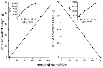
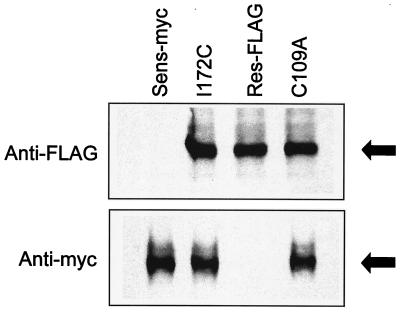
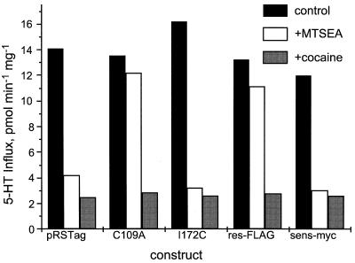
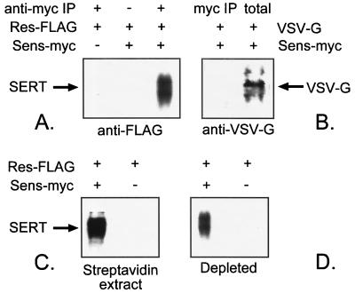
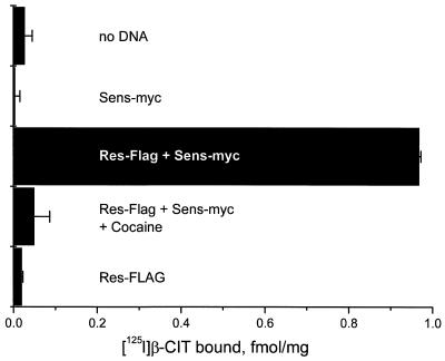
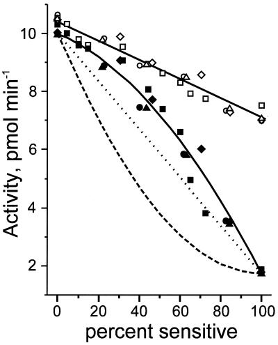
Two forms of serotonin transporter (SERT) were prepared with different epitope tags. When co-expressed in HeLa cells, the form containing a FLAG tag (Res-FLAG) was associated with the form containing a c-myc tag (Sens-myc). Antibody against c-myc precipitated Res-FLAG from detergent extracts of cells expressing both forms, but not when Res-FLAG was expressed alone. The specificity of the interaction was demonstrated by the observation that anti-myc antibodies did not precipitate the unrelated vesicular stomatitis virus coat glycoprotein when it was co-expressed with Sens-myc. Sens-myc contained a reactive cysteine at position 172, which reacted with both (2-aminoethyl)methanethiosulfonate and N-biotinylaminoethyl methanethiosulfonate on the surface of intact cells. Sens-myc, but not Res-FLAG, was inactivated by these reagents. When co-expressed with Sens-myc, functionally active Res-FLAG was precipitated by immobilized streptavidin from digitonin-solubilized cells that had been treated with N-biotinylaminoethyl methanethiosulfonate. In cells co-expressing mixtures of Sens-myc and Res-FLAG, the amount of inactivation by (2-aminoethyl)methanethiosulfonate was less than expected if the two forms were independent. The results are consistent with a dimeric form of SERT with functional interactions between subunits, and with association of dimers into a higher order complex, possibly a tetramer.


Figures

Figure 1
Figure 1
Standard curves for quantitation of SERT mutant expression. The indicated amounts of cell protein from cells expressing SERT C109A containing both c-myc and FLAG epitope tags were separated by PAGE and were visualized by Western blot analysis, and the integrated density values were determined by densitometry. The relationship between C109A lysate applied and the integrated density value (IDV) is shown in the insets for c-myc (Left) and FLAG (Right) antibodies. Lysates from cells expressing mixtures of Res-FLAG and Sens-myc were separated on the same gels, and the equivalent amount of C109A was determined from the C109A standard curve for each mixture.
Figure 2
Figure 2
Antigenic reactivity of SERT mutants. Each panel represents a Western blot of HeLa cells expressing either Res-FLAG, which contains FLAG, but not myc epitopes, Sens-myc, which contains myc but not FLAG, C109A, or I172C, each of which contain both tags. The upper blot was visualized with antibody against FLAG and the lower blot with antibody against c-myc.
Figure 3
Figure 3
Sensitivity of SERT mutants to MTSEA. HeLa cells expressing either pRSTag (SERT wild type), C109A, I172C, Res-FLAG, or Sens-myc were incubated with 0.25 mM MTSEA for 10 min and then were assayed for transport activity. Filled bars, control; open bars, MTSEA; gray bars, cocaine control for nonspecific activity.
Figure 4
Figure 4
Co-precipitation of Res-FLAG with Sens-myc. (A) HeLa cells transfected with equal amounts of Res-FLAG and Sens-myc cDNA, or with Res-FLAG alone, were solubilized and treated with Protein-A beads and, where indicated, antibody against c-myc. The immunoprecipitates were separated by SDS/PAGE and were blotted with anti-FLAG antibody as described. (B) Cells expressing Sens-myc and VSV-G protein were treated as in A. The immunoprecipitate in the left lane is compared with the initial cell lysate in the right lane. (C) Sens-myc on the surface of cells expressing Res-FLAG and Sens-myc, or Res-FLAG alone, was labeled with 1 mM MTSEA-biotin, solubilized, precipitated with streptavidin-agarose, and Western blotted with anti-FLAG antibody. (D) The remaining soluble extract after depletion of cell lysate with MTSEA-biotin, and streptavidin-agarose, as in C, was immunoprecipitated and Western blotted as in A. Although the sample in C represents quantitative precipitation of biotinylated proteins, the fraction of intracellular SERT precipitated with anti-myc antibodies (D) was not determined.
Figure 5
Figure 5
Precipitation of active Res-FLAG in digitonin by biotinylated Sens-myc. HeLa cells (500 μg of cell protein) transfected with equal amounts of Res-FLAG and Sens-myc cDNA, or with Res-FLAG or Sens-myc alone, were treated with 1 mM MTSEA-biotin for 10 min, were washed, and were solubilized in 0.5 ml of binding buffer (18) containing 1% digitonin. The lysate was incubated with 0.5 ml of packed streptavidin-agarose beads for 2 h at 4°C, and the beads were washed three times with the same buffer containing 0.06% digitonin. Each sample of packed beads was incubated with 500 μl of 0.03 nM 2β-carbomethoxy-3β-(4-[125I]iodophenyl)tropane ([125I]β-CIT) in binding buffer for 30 min at 25°C in the presence (where indicated) of 100 μM cocaine, and the beads were then collected on #32 glass-fiber filters (Schleicher & Schuell), were washed, and were counted.
Figure 6
Figure 6
Inactivation of Res-FLAG and Sens-myc mixtures by MTSEA. HeLa cells were transfected with various amounts of Res-FLAG and Sens-myc cDNA and were assayed for 5-HT transport. The percent of Sens-myc relative to total SERT was determined by quantitative immunoblotting and is indicated on the abscissa. Four separate experiments are shown, with separate symbols (squares, circles, triangles, and diamonds) for each experiment. The experiment shown by the squares was normalized so that the activity of 100% Res-FLAG matched the other three experiments. Filled symbols represent rates of transport after treatment with 0.25 mM MTSEA for 10 min. Three lines were plotted according to predictions of the amount of activity remaining after MTSEA treatment. The dotted line is the activity expected if no interaction occurred between Res-FLAG and Sens-myc, and the amount of inactivation was equal to the amount of activity contributed by Sens-myc. The dashed line assumes random association of Res-FLAG and Sens-myc into dimers and that all of the activity of all dimers containing Sens-myc was sensitive to MTSEA. The lower solid line assumes random dimer formation and also that only dimers containing two Sens-myc subunits would be sensitive to MTSEA. In this model, no activity would be lost from dimers containing one subunit each of Res-FLAG and Sens-myc.

References

    1. Miller C. Science. 1992;258:240–241. - PubMed
    1. Mitchell P. Res Microbiol. 1990;141:286–289. - PubMed
    1. Eskandari S, Wright E M, Kreman M, Starace D M, Zampighi G A. Proc Natl Acad Sci USA. 1998;95:11235–11240. - PMC - PubMed
    1. Sahin-Toth M, Lawrence M C, Kaback H R. Proc Natl Acad Sci USA. 1994;91:5421–5425. - PMC - PubMed
    1. Hackenberg H, Klingenberg M. Biochemistry. 1980;19:548–555. - PubMed

Publication types

LinkOut - more resources