. 2000 Mar 28;97(7):3314-8.
doi: 10.1073/pnas.97.7.3314.

Reciprocal domain evolution within a transactivator in a restricted sequence space

Affiliations

Reciprocal domain evolution within a transactivator in a restricted sequence space

K Juarez et al. Proc Natl Acad Sci U S A. .

Abstract

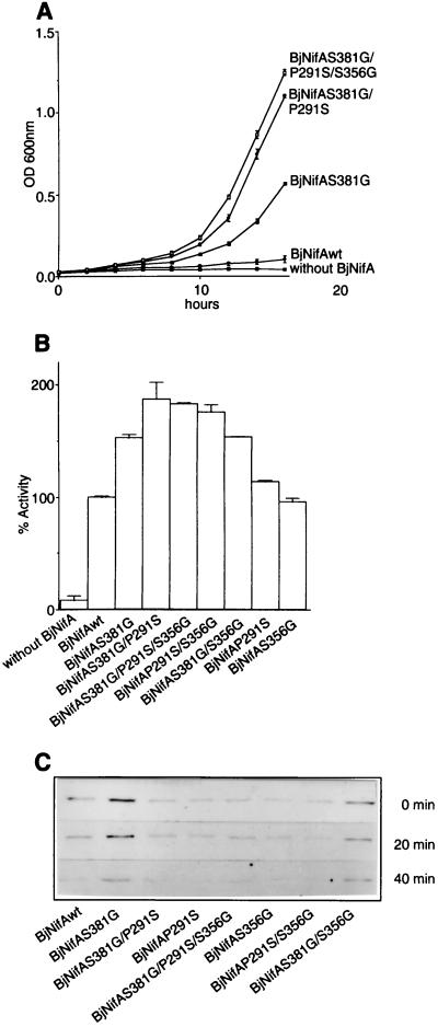
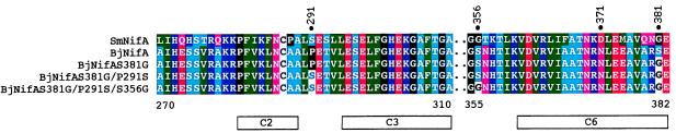
offhough the concept of domain merging and shuffling as a major force in protein evolution is well established, it has been difficult to demonstrate how domains coadapt. Here we show evidence of coevolution of the Sinorhizobium meliloti NifA (SmNifA) domains. We found that, because of the lack of a conserved glycine in its DNA-binding domain, this transactivator protein interacts weakly with the enhancers. This defect, however, was compensated by evolving a highly efficient activation domain that, contrasting to Bradyrhizobium japonicum NifA (BjNifA), can activate in trans. To explore paths that lead to this enhanced activity, we mutagenized BjNifA. After three cycles of mutagenesis and selection, a highly active derivative was obtained. Strikingly, all mutations changed to amino acids already present in SmNifA. Our artificial process thus recreated the natural evolution followed by this protein and suggests that NifA is trapped in a restricted sequence space with very limited solutions for higher activity by point mutation.


Figures

Figure 1
Figure 1
DNA-binding properties of different NifA proteins and amino acid sequence of various hth motifs. (A) In vivo DMS footprinting of the K. pneumoniae nifH enhancer with different NifA proteins, as indicated. Protection from methylation of guanine-136 by KpNifA and BjNifA is indicated. This residue is part of the TGT-N10-ACA nifH enhancer. (B) Sequence alignment of the hth motif of several NifA and the SmNifA and BjNifA mutant proteins constructed in this work. The SwissProt name for each protein is indicated. (C) Alignment of the hth motifs of proteins whose structures have been solved as cocrystals with their DNA-binding sites. SwissProt (first column) and Protein Data Bank (last column) names are specified. 6CRO, lambda cro; 3CRO, 434 cro; 1LMB, lambda repressor; 1TRO, trp repressor; 1LCC, lac repressor; 1CGP, catabolite activator protein (cap); 2ORI, 434 repressor; 1HDD, Drosophila homeodomain protein; 1HCR, Hin recombinase. A, Ala; C, Cys; D, Asp; E, Glu; F, Phe; G, Gly; H, His; I, Ile; K, Lys; L, Leu; M, Met; N, Asn; P, Pro; Q, Gln; R, Arg; S, Ser; T, Thr; V, Val; W, Trp; and Y, Tyr.
Figure 2
Figure 2
Analysis of DNA binding, protein stability, and transcriptional activation of SmNifA, BjNifA, and their mutant derivatives. (A) Footprinting in vivo of the K. pneumoniae nifH enhancer with different NifA proteins and mutant derivatives, as indicated. Guanine-136 of the nifH enhancer is marked. (B and C) Immunodetection of the SmNifA and BjNifA (see Materials and Methods) proteins and their mutant derivatives. Antibodies raised against synthetic peptides corresponding to the hth of SmNifA or BjNifA were used for the immunodetection shown in B and C, respectively. Mr values of control proteins are denoted. Note that the anti-SmNifA hth peptide antibodies did not react against either BjNifA or SmNifAE10G/K11L and only weakly against SmNifAE10G, but efficiently recognized BjNifAG10E/L11K. Conversely, antibodies raised against BjNifA hth peptide did not recognize either SmNifA or BjNifAG10E/L11K and reacted weakly against SmNifAE10G, but efficiently recognized SmNifAE10G/K11L. (D and E) Transcriptional activation of a nifH-lacZ fusion carrying (solid bars) or deleted of (open bars) the enhancer by different NifA proteins and their mutant derivatives, as indicated. Because SmNifA and BjNifA are coded in different vectors and expressed from different promoters, it is not feasible to correlate actual β-galactosidase activities. Values represent the mean ± SD of three independent experiments and are expressed as percentage of β-galactosidase activity in Miller units.
Figure 3
Figure 3
Transcriptional activity, stability, and sensitivity to oxygen of the BjNifA derivatives. (A) Growth curves of strains carrying the wild type or the directly evolved BjNifA derivatives, as indicated. Cells were grown on NFDM medium with Cm (40 μg/ml) under aerobic conditions. The level of expression of the cat gene in pVB007, under the S. meliloti nifH promoter (deleted of the enhancer), and, therefore, the resistance to Cm is dependent on the activity of each BjNifA derivative. Values represent the mean ± SD of three independent experiments. (B) Transcriptional activation of a nifH-lacZ fusion by the BjNifA mutant derivatives. (C) Stability and sensitivity of BjNifA mutant proteins to oxygen. To analyze whether the mutations affected the stability of the protein or any intrinsic function leading to a higher transcriptional activation, we grew strains carrying each of the mutant BjNifA proteins in microaerobic cultures, and the amount of the protein was detected after being shifted to heavily aerated flasks. Samples were taken at time 0 min, 20 min, and 40 min, and the BjNifA derivatives were immunodetected in soluble cell extracts with an antibody raised against a polypeptide comprising part of the central domain. All mutant proteins had similar stability and oxygen sensitivity because they decreased at the same rate as the wild type after being shifted to air.
Figure 4
Figure 4
Multiple sequence alignment of SmNifA, BjNifA, and its mutant derivatives. Only regions near the mutated residues on BjNifA are shown. Dots indicate discontinuity of the alignment. Numbers above the sequences indicate amino acid positions mutated in BjNifA. Numbers below the alignment show amino acid positions of BjNifA. Boxes indicate the highly conserved regions in the EBP proteins (9, 10). The central domains of BjNifA and SmNifA are 68% identical, whereas the degree of identity of the whole family is around 40%.

References

    1. Henikoff E S, Greene A, Pietrokovski S, Bork P, Attwood T K, Hood L. Science. 1997;278:609–614. - PubMed
    1. Doolittle R F. Annu Rev Biochem. 1995;64:287–314. - PubMed
    1. Doolittle R F, Bork P. Sci Am. 1993;269:50–56. - PubMed
    1. Netzer W J, Hart F U. Nature (London) 1997;388:343–349. - PubMed
    1. Chothia C. Nature (London) 1992;357:543–544. - PubMed

Publication types

MeSH terms

LinkOut - more resources