. 2000 Mar 28;97(7):3062-6.
doi: 10.1073/pnas.97.7.3062.

A method to identify cDNAs based on localization of green fluorescent protein fusion products

Affiliations

A method to identify cDNAs based on localization of green fluorescent protein fusion products

K Misawa et al. Proc Natl Acad Sci U S A. .

Abstract

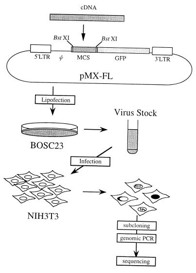
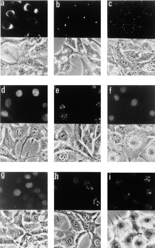
We previously established a high-efficiency, retrovirus-mediated expression cloning method. Using this system, we now have developed an expression cloning method (FL-REX; fluorescence localization-based retrovirus-mediated expression cloning) in which cDNAs can be isolated based on the subcellular localization of their protein products. Complementary DNAs generated from mRNA using random hexamers were fused to the cDNA of green fluorescent protein (GFP) in the pMX retrovirus vector. The resulting cDNA-GFP fusion library was transfected into retrovirus-packaging cells, and the derived retroviruses were used to infect NIH 3T3 cells. Infected cells then were screened to identify cDNAs of interest through the subcellular localization of the GFP-fusion products. Using FL-REX, we have identified 25 cDNAs, most of which showed reasonable subcellular localization as GFP-fusion proteins, indicating that FL-REX is useful for identification of proteins that show specific intracellular localization.


Figures

Figure 1
Figure 1
A rationale of FL-REX. A retrovirus vector pMX-FL was designed for the construction of the cDNA-GFP fusion library; cDNAs can be inserted into BstXI sites for the fusion with the GFP cDNA. After converting the library to retroviruses by using Bosc23-packaging cells, the derived retroviruses were used to infect NIH 3T3 cells, and the clones of interest were subjected for further analysis as described in the experimental protocol. LTR, long terminal repeat; Ψ, retrovirus-packaging signal; MCS, multicloning site.
Figure 2
Figure 2
Photographs of the clones with specific intercellular localization patterns. The upper part of each photo shows GFP fluorescence, and the lower part shows corresponding phase-contrast images. The lower part of b is the combination of both images. (a) Clone 1, vimentin (fibrous). (b) Clone 2, mouse homologue of human R27216–1 (perinuclear spotty). (c) Clone 5, a novel gene related to keratin family (cytoplasmic speckle). (d) Clone 10, mouse ZNF207 (nuclear). (e) Clone 24, nucleophosmin (nucleolar). (f) Clone 17, hnRNP C (nuclear spotty). (g) Clone 18, mouse SET (nuclear speckle). (h) Clone 20, mouse PML (nuclear speckle). (i) Clone 21, a novel gene (nuclear speckle).

References

    1. Kitamura T, Onishi M, Kinoshita S, Shibuya A, Miyajima A, Nolan G P. Proc Natl Acad Sci USA. 1995;92:9146–9150. - PMC - PubMed
    1. Whitehead I, Kirk H, Kay R. Mol Cell Biol. 1995;15:704–710. - PMC - PubMed
    1. Rayner J R, Gonda T J. Mol Cell Biol. 1994;14:880–887. - PMC - PubMed
    1. Deng H K, Unutmaz H K, Kewal Ramani V N, Littman D R. Nature (London) 1997;388:296–300. - PubMed
    1. Hitoshi Y, Lorens J, Kitada S, Fisher J, LaBarge M, Ring H Z, Francke U, Reed J C, Kinoshita S, Nolan G P. Immunity. 1998;8:461–471. - PubMed

Publication types

Associated data