. 2000 Mar 28;97(7):3142-7.
doi: 10.1073/pnas.97.7.3142.

Recognition of a conserved class of RNA tetraloops by Saccharomyces cerevisiae RNase III

Affiliations

Recognition of a conserved class of RNA tetraloops by Saccharomyces cerevisiae RNase III

G Chanfreau et al. Proc Natl Acad Sci U S A. .

Abstract

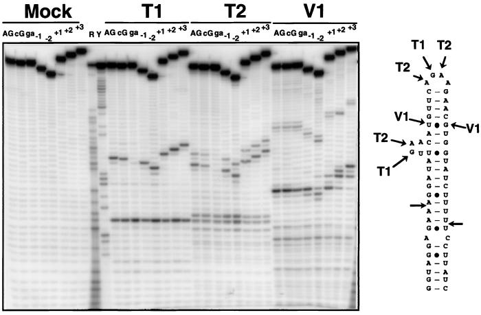
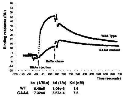
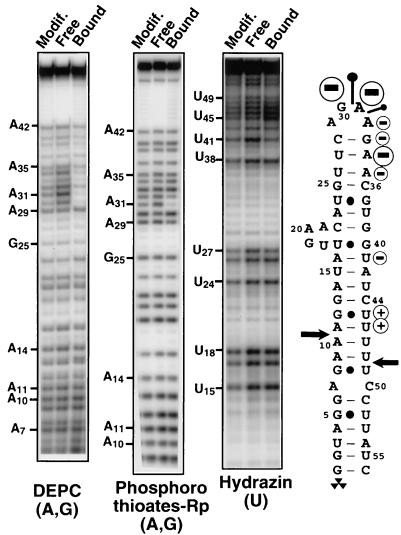
Ribonucleases III are double-stranded RNA (dsRNA) endonucleases required for the processing of a large number of prokaryotic and eukaryotic transcripts. Although the specificity of bacterial RNase III cleavage relies on antideterminants in the dsRNA, the molecular basis of eukaryotic RNase III specificity is unknown. All substrates of yeast RNase III (Rnt1p) are capped by terminal tetraloops showing the consensus AGNN and located within 13-16 bp to Rnt1p cleavage sites. We show that these tetraloops are essential for Rnt1p cleavage and that the distance to the tetraloop is the primary determinant of cleavage site selection. The presence of AGNN tetraloops also enhances Rnt1p binding, as shown by surface plasmon resonance monitoring and modification interference studies. These results define a paradigm of RNA loops and show that yeast RNase III behaves as a helical RNA ruler that recognizes these tetraloops and cleaves the dsRNA at a fixed distance to this RNA structure. These results also indicate that proteins belonging to the same class of RNA endonucleases require different structural elements for RNA cleavage.


Figures

Figure 1
Figure 1
Single-turnover cleavage of wild-type and mutant model substrates by recombinant Rnt1p. 5′ end-labeled RNAs (20 fmol) were incubated for 2 min with recombinant His6–Rnt1p (800 fmol) or in buffer with no enzyme (Mock), and products were fractionated on a 10% sequencing gel. The arrows labeled WT, −1, −2, +1, +2, and +3 indicate the positions of the major cleavage products for the wild-type substrate and for the corresponding insertion-deletion mutants, respectively. The radioactive symbol indicates the 5′ end label of the RNA.
Figure 2
Figure 2
RNase probing of 5′ end-labeled wild-type and mutant model substrates. Shown are the cleavage products of wild-type and various mutant substrates with RNase T1, T2, and V1 or with buffer alone (Mock). The cleavage sites of these RNases on the wild-type substrate are indicated on the secondary structure. R and Y are size markers obtained by iodine cleavage of RNAs substituted with purine (R) or pyrimidine (Y) phosphorothioates. AG, wild-type substrate; cG, CGAA tetraloop mutant; ga, GAAA tetraloop mutant.
Figure 3
Figure 3
Binding of wild-type (WT) and mutant snR47 substrates by immobilized Rnt1p. The binding and the dissociation of the RNAs were followed by monitoring the response (RU, arbitrary units) on a BIAcore system. Shown is a representative profile for the wild-type and the GAAA mutant substrates at a concentration of 50 nM. The association rate ka and dissociation rate kd are indicated, as well as the resulting dissociation constant Kd for both RNAs. The ka reported in the figure is the apparent second order rate constant (M−1⋅s−1) and depends on the RNA concentration, whereas the kd was independent of RNA concentration.
Figure 4
Figure 4
Interference of DEPC, phosphorothioate, and hydrazine modifications of the snR47 substrate on Rnt1p binding. RNAs modified by DEPC, hydrazine, or by incorporation of purine phosphorothioates (thios A and G) were incubated with recombinant Rnt1p protein, and bound and free RNAs were gel purified. After cleavage of DEPC- or hydrazine-modified RNAs by aniline and of phosphorothioate-modified RNAs by iodine, the corresponding cleaved RNAs populations were fractionated on 12% polyacrylamide gels. The strongest effects of modifications are indicated on the secondary structure of the snR47 stem loop. “−” and “+” signs indicate inhibitory and stimulatory effects of base modifications on Rnt1p binding, respectively. Pins indicate the location of the nonbridging Rp phosphate oxygens where substitution by sulfur inhibits Rnt1p binding. The size of the circles and of the pins is roughly proportional to the inhibitory effect, as quantitated by PhosphorImager analysis. No inhibitory effects of phosphorothioate incorporation at pyrimidines were found (data not shown). Cytosines and the 5′ first 5 nt of the substrate were not mapped.

References

    1. Robertson H D, Webster R E, Zinder N D. J Biol Chem. 1968;243:82–91. - PubMed
    1. Court D. In: Control of mRNA Stability. Brawerman G, Belasco J, editors. New York: Academic; 1993. pp. 70–116.
    1. Apirion D, Miczak A. BioEssays. 1993;15:113–120. - PubMed
    1. Nicholson A W. In: Ribonucleases: Structures and Functions. D'Alessio G, Riordan J F, editors. New York: Academic; 1997. pp. 1–49.
    1. Zhang K, Nicholson A W. Proc Natl Acad Sci USA. 1997;94:13437–13441. - PMC - PubMed

LinkOut - more resources