. 2000 Mar 1;14(5):549-58.

An auxiliary mode of apoptotic DNA fragmentation provided by phagocytes

Affiliations

An auxiliary mode of apoptotic DNA fragmentation provided by phagocytes

D McIlroy et al. Genes Dev. .

Abstract

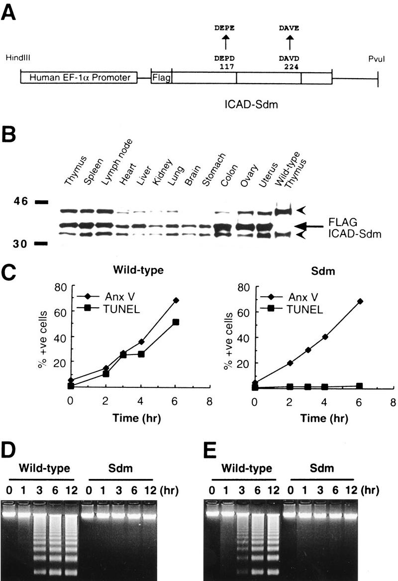
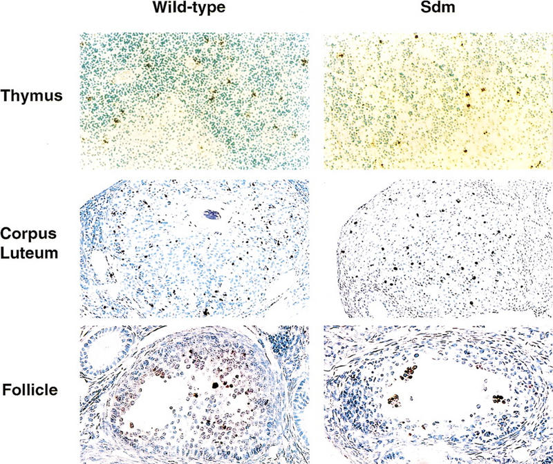
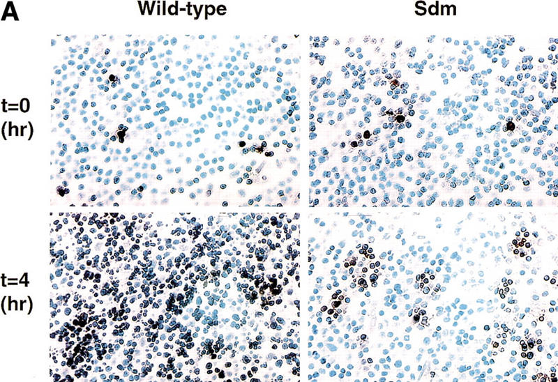
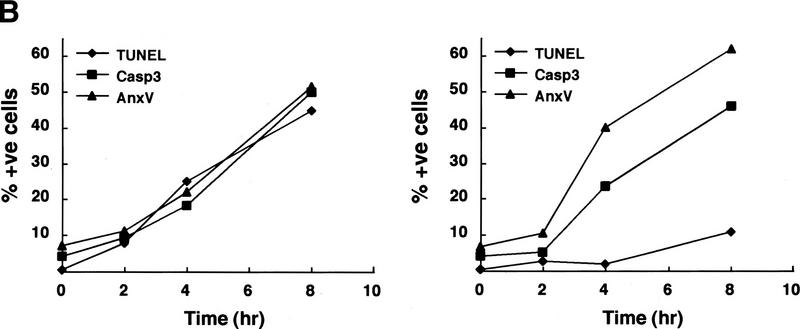
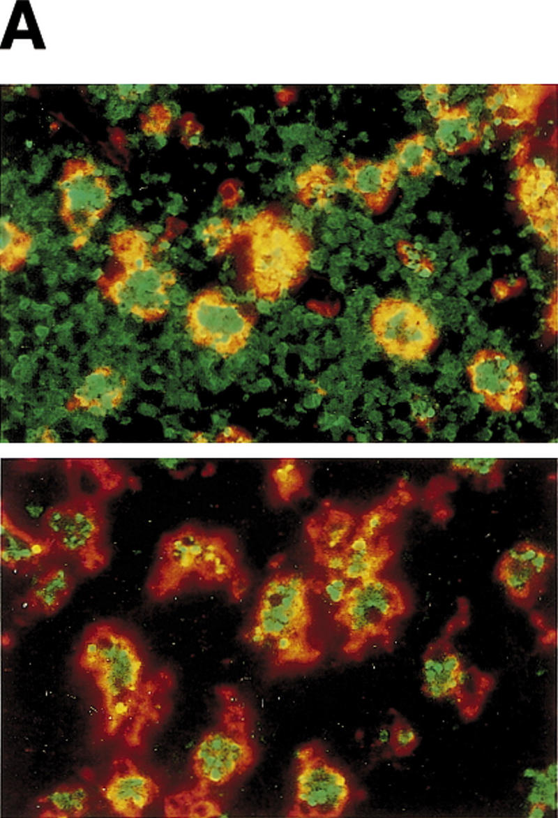
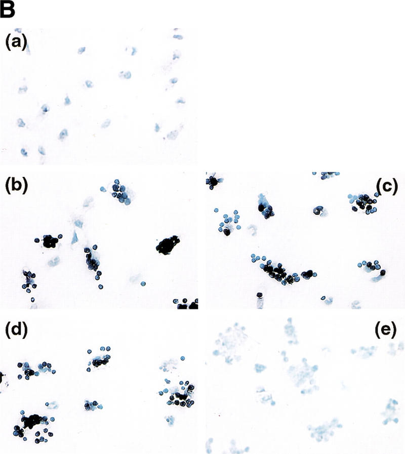
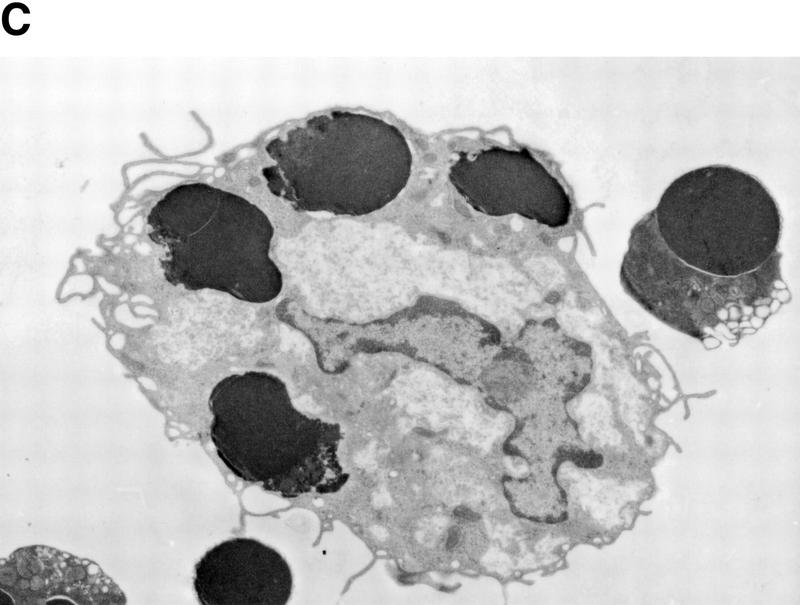
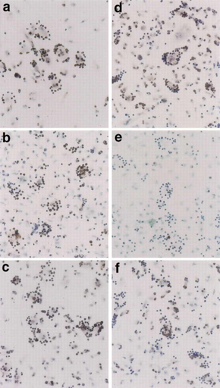
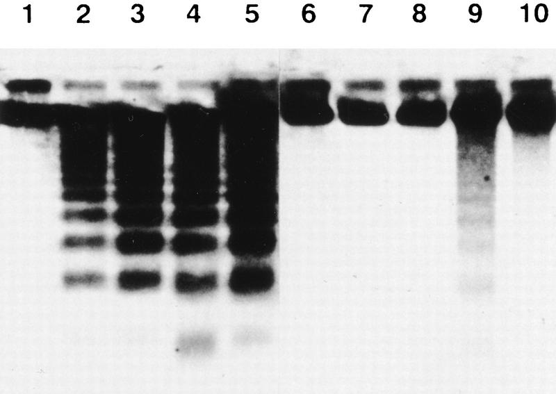
CAD (caspase-activated DNase) can cause DNA fragmentation in apoptotic cells. Transgenic mice that ubiquitously express a caspase-resistant form of the CAD inhibitor (ICAD) were generated. Thymocytes prepared from the mice were resistant to DNA fragmentation induced by a variety of stimuli. However, similar numbers of TUNEL-positive cells were present in adult tissues of transgenic and wild-type mice. Exposure to gamma-irradiation caused a striking increase in the number of TUNEL-positive cells in the thymus of wild-type, but not transgenic, mice. TUNEL-positive nuclei in transgenic mice were confined to thymic macrophages. When apoptotic thymocytes from the transgenic mice were cocultured with macrophages, the thymocytes underwent phagocytosis and their chromosomal DNA underwent fragmentation. This DNA fragmentation was sensitive to inhibitors that block the acidification of lysosomes. Hence, we conclude that the DNA fragmentation that occurs during apoptosis not only can result cell-autonomously from CAD activity but can also be attributed to a lysosomal acid DNase(s), most likely DNase II, after the apoptotic cells are engulfed.


Figures

Figure 1
Figure 1
Ubiquitous expression of caspase-resistant ICAD-S in transgenic mice and its effect on in vitro apoptosis. (A) A schematic view of the expression plasmid used to produce the ICAD–Sdm transgenic mice. A 0.8-kb ICAD-S cDNA (open boxes) carrying the D117E and D224E mutations was placed under the control of a 2.4-kb DNA fragment that carries the 5′-flanking sequence, exon 1, and intron 1 of the human EF-1α gene. (B) Ubiquitous expression of the ICAD–Sdm protein. The cell lysates were prepared from the indicated tissues of the transgenic mice and the thymus of wild-type mice. The lysates (20-μg protein) were analyzed by Western blotting with a rabbit antibody against mouse ICAD. The Flag-tagged ICAD–Sdm is indicated by an arrow; arrowheads represent the endogenous ICAD-L and ICAD-S. (C) No DNA degradation in the transgenic thymocytes by γ-irradiation. Mouse thymocytes from the ICAD–Sdm transgenic (right) or wild-type (left) littermate mice were exposed for 10 min to γ-rays (1.2 Gy/min) and cultured for the indicated periods of time. Cells were then stained with FITC-labeled annexin V for apoptotic cells (♦) or with ApopTag fluorescein for TUNEL-positive cells (█) and analyzed by flow cytometry. The results are plotted as percentages of total cells. (D-E) No DNA fragmentation in the transgenic thymocytes by treatment with dexamethasone or anti-Fas antibody. Mouse thymocytes (1 × 107 cells) from the ICAD–Sdm transgenic mice or wild-type littermates were treated for the indicated periods of time with either 10 μm dexamethasone (D) or 1 μg/ml of anti-Fas antibody in the presence of 30 μm cycloheximide (E). The chromosomal DNA was analyzed by electrophoresis on an agarose gel.
Figure 2
Figure 2
TUNEL-positive cells in various tissues of wild-type and ICAD–Sdm transgenic mice. Paraffin sections were prepared from the thymus and ovary of adult wild-type or ICAD–Sdm transgenic mice. TUNEL-positive cells were detected using an Apotag kit, and DAB-black as a peroxidase substrate. Nuclei were counterstained with hematoxylin. Original magnifications: thymus, 100×; corpus luteum, 100×; ovarian follicules, 150×.
Figure 3
Figure 3
Effect of γ-irradiation on TUNEL-positive cells in the thymus. (A) In situ detection of TUNEL-positive cells in the thymus. Wild-type and ICAD–Sdm transgenic mice were left untreated or exposed for 11 min to γ-rays (1.2 Gy/min). After 4 hr, the mice were sacrificed, and paraffin sections of the thymic cortex were immunohistochemically stained for TUNEL reaction, using an Apotag kit. The sections were then counterstained with methyl green. Original magnifications, 400×. (B) Apoptosis of thymocytes after γ-irradiation. Wild-type (left) and ICAD–Sdm (right) mice were irradiated as above. Thymocytes were prepared from the irradiated mice at the indicated time. Aliquots of 1 × 105 cells were stained with FITC-conjugated annexin V (▴), ApopTag-fluorescein for TUNEL-positive cells (♦), or PhiPhiLux-G1D2 for the activated caspase 3 (█), and analyzed by flow cytometry. The results are expressed as percentages of total cells.
Figure 3
Figure 3
Effect of γ-irradiation on TUNEL-positive cells in the thymus. (A) In situ detection of TUNEL-positive cells in the thymus. Wild-type and ICAD–Sdm transgenic mice were left untreated or exposed for 11 min to γ-rays (1.2 Gy/min). After 4 hr, the mice were sacrificed, and paraffin sections of the thymic cortex were immunohistochemically stained for TUNEL reaction, using an Apotag kit. The sections were then counterstained with methyl green. Original magnifications, 400×. (B) Apoptosis of thymocytes after γ-irradiation. Wild-type (left) and ICAD–Sdm (right) mice were irradiated as above. Thymocytes were prepared from the irradiated mice at the indicated time. Aliquots of 1 × 105 cells were stained with FITC-conjugated annexin V (▴), ApopTag-fluorescein for TUNEL-positive cells (♦), or PhiPhiLux-G1D2 for the activated caspase 3 (█), and analyzed by flow cytometry. The results are expressed as percentages of total cells.
Figure 4
Figure 4
TUNEL-positive thymocytes in macrophages. (A) Ingestion of TUNEL-positive cells in thymuses of ICAD–Sdm transgenic mice by macrophages. Wild-type (top) and ICAD–Sdm transgenic mice (bottom) were exposed for 11 min to γ-rays (1.2 Gy/min). Four hours later, cryosections of the thymus were double-stained for TUNEL-positive cells (green) and F4/80 antigen (red). Original magnifications, 400×. (B) In vitro generation of TUNEL-positive cells by macrophages. Thioglycollate-elicited peritoneal macrophages were resuspended in RPMI medium containing 10% FCS, distributed in eight-well glass chamber slides, and cultured overnight at 37°C. Mouse thymocytes from wild-type (b,d) or ICAD–Sdm (c,e) mice were incubated at 37°C for 3.5 hr with 10 μm dexamethasone in RPMI medium containing 10% FCS. Apoptotic thymocytes were then added to macrophage cultures, and phagocytosis was allowed to proceed for 1 hr in the absence (b,c) or presence (d,e) of 100 μm chloroquine. Cells not undergoing phagocytosis were removed, and the adherent macrophages were fixed with 1% paraformaldehyde, and stained using the TUNEL reaction, and lightly counterstained with Wright's stain. As a control, macrophages without thymocytes were subjected to the TUNEL staining procedure (a). Original magnifications, 400×. (C) Electron micrograph of macrophages carrying thymocytes that underwent phagocytosis. The apoptotic thymocytes from wild-type mice underwent phagocytosis by macrophages as described above. The cells were fixed, stained with uranyl acetate, and examined using a Hitachi H7100 transmission electron microscope.
Figure 4
Figure 4
TUNEL-positive thymocytes in macrophages. (A) Ingestion of TUNEL-positive cells in thymuses of ICAD–Sdm transgenic mice by macrophages. Wild-type (top) and ICAD–Sdm transgenic mice (bottom) were exposed for 11 min to γ-rays (1.2 Gy/min). Four hours later, cryosections of the thymus were double-stained for TUNEL-positive cells (green) and F4/80 antigen (red). Original magnifications, 400×. (B) In vitro generation of TUNEL-positive cells by macrophages. Thioglycollate-elicited peritoneal macrophages were resuspended in RPMI medium containing 10% FCS, distributed in eight-well glass chamber slides, and cultured overnight at 37°C. Mouse thymocytes from wild-type (b,d) or ICAD–Sdm (c,e) mice were incubated at 37°C for 3.5 hr with 10 μm dexamethasone in RPMI medium containing 10% FCS. Apoptotic thymocytes were then added to macrophage cultures, and phagocytosis was allowed to proceed for 1 hr in the absence (b,c) or presence (d,e) of 100 μm chloroquine. Cells not undergoing phagocytosis were removed, and the adherent macrophages were fixed with 1% paraformaldehyde, and stained using the TUNEL reaction, and lightly counterstained with Wright's stain. As a control, macrophages without thymocytes were subjected to the TUNEL staining procedure (a). Original magnifications, 400×. (C) Electron micrograph of macrophages carrying thymocytes that underwent phagocytosis. The apoptotic thymocytes from wild-type mice underwent phagocytosis by macrophages as described above. The cells were fixed, stained with uranyl acetate, and examined using a Hitachi H7100 transmission electron microscope.
Figure 5
Figure 5
Effect of bafilomycin on macrophage-induced generation of TUNEL-positive cells. Thymocytes from wild-type (ac) or ICAD–Sdm (df) mice were incubated at 37°C for 3.5 hr with 10 μm dexamethasone in RPMI medium containing 10% FCS. Bafilomycin A1 at 50 nm was added to the culture for the last hour of incubation in c and f. Thioglycollate-elicited peritoneal macrophages were resuspended in RPMI medium containing 10% FCS, distributed in eight-well glass chamber slides, cultured overnight, and incubated at 37°C for 1 hr in the presence (b,e) or absence (a,c,d,e) of 50 nm bafilomycin A1. Thymocytes and macrophages were washed to remove bafilomycin, cocultured at 37°C for 1 hr, and stained using the TUNEL reaction as described in Materials and Methods. Original magnification, 400×.
Figure 6
Figure 6
DNA fragmentation in thymocytes after phagocytosis by macrophages. Wild-type (lanes 15) and ICAD–Sdm transgenic (lanes 610) mice were given BrdU for 7 days in drinking water, and their thymocytes were prepared. The thymocytes were left untreated (lanes 1,6) or treated (lanes 25,710) with 10 μm dexamethasone as described above. The apoptotic thymocytes were then added to the culture of peritoneal macrophages from wild-type B6 mice, and incubated at 37°C for 1 hr in the absence (lanes 3,4,8,9) or presence (lanes 5,10) of 100 μm chloroquine. DNA was prepared from nonadherent (lanes 3,8) or adherent (lanes 4,5,9,10) cells, separated by electrophoresis on an agarose gel, transferred to a Hybond-N membrane, and probed with a peroxidase-conjugated anti-BrdU monoclonal antibody. Bands were revealed by chemiluminescence.

References

    1. Adachi M, Suematsu S, Kondo T, Ogasawara J, Tanaka T, Yoshida N, Nagata S. Targeted mutation in the Fas gene causes hyperplasia in the peripheral lymphoid organs and liver. Nat Genet. 1995;11:294–300. - PubMed
    1. Aliprantis AO, Diez-Roux G, Mulder LC, Zychlinsky A, Lang RA. Do macrophages kill through apoptosis? Immunol Today. 1996;17:573–576. - PubMed
    1. Ashkenazi A, Dixit VM. Death receptors: Signaling and modulation. Science. 1998;281:1305–1308. - PubMed
    1. Austyn JM, Gordon S. F4/80, a monoclonal antibody directed specifically against the mouse macrophage. Eur J Immunol. 1981;11:805–815. - PubMed
    1. Baker KP, Baron WF, Henzel WJ, Spencer SA. Molecular cloning and characterization of human and murine DNase II. Gene. 1998;215:281–289. - PubMed

Publication types

MeSH terms

LinkOut - more resources