. 2000 Mar 1;14(5):585-95.

WISP-1 is a Wnt-1- and beta-catenin-responsive oncogene

Affiliations

WISP-1 is a Wnt-1- and beta-catenin-responsive oncogene

L Xu et al. Genes Dev. .

Abstract

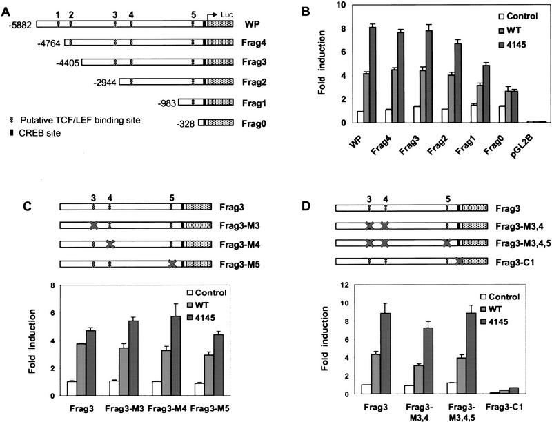
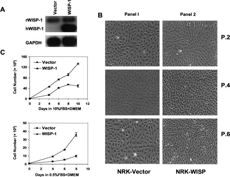
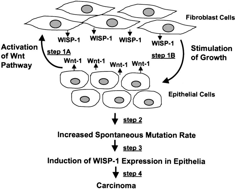
WISP-1 (Wnt-1 induced secreted protein 1) is a member of the CCN family of growth factors. This study identifies WISP-1 as a beta-catenin-regulated gene that can contribute to tumorigenesis. The promoter of WISP-1 was cloned and shown to be activated by both Wnt-1 and beta-catenin expression. TCF/LEF sites played a minor role, whereas the CREB site played an important role in this transcriptional activation. WISP-1 demonstrated oncogenic activities; overexpression of WISP-1 in normal rat kidney fibroblast cells (NRK-49F) induced morphological transformation, accelerated cell growth, and enhanced saturation density. Although these cells did not acquire anchorage-independent growth in soft agar, they readily formed tumors in nude mice, suggesting that appropriate cellular attachment is important for signaling oncogenic events downstream of WISP-1.


Figures

Figure 1
Figure 1
(A) Schematic of β-catenin mutants. (Stippled boxes) The armadillo repeats. (B) Immunoprecipitation of β-catenin mutants in C57MG cells. Myc-tagged β-catenin mutants were transfected into C57MG cells. Cells were then metabolically labeled with 35S-methionine for 5 hr and transfected β-catenin was immunoprecipitated with anti-myc antibody 9E10 (CalBiochem). Signals were visualized by autoradiography. (C) Mutant NLSs localized in the nucleus instead of cytoplasm. Mutant 4145 (a) and mutant NLS (b) were transfected into 293 cells. Immunofluorescence staining was performed with anti-myc antibody as the first antibody, followed by sequential incubation with biotinylated goat anti-mouse IgG and FITC-conjugated streptavidin (Jackson Immunoresearch Laboratories). (D) Response of TopFlash reporter to β-catenin mutants. The 293 cell were transfected with 0.5 μg of TopFlash reporter together with 0.5 μg of empty pCS2/MT vector, or different β-catenin mutants. A total of 1 μg of pCS2/MT plasmid DNA was used to make up the total DNA to 2 μg; 0.1 μg of CMV–β-gal was cotranfected to normalize transfection efficiency.
Figure 2
Figure 2
(A) Northern blot analysis of WISP-1 RNA in C57MG cells expressing mutant β-catenin or Wnt-1. Total RNA (10 μg) from the cells was loaded onto each lane. The blot was hybridized with a full-length mouse WISP-1 probe. For loading control, the blot was rehybridized with a probe for mouse GAPDH gene. (B) Response of the WISP-1 promoter luciferase reporter (pGL2B-WP, WISP-1 sequence −5882 ∼−42, translation start site as +1) to Wnt-1. C57MG cells were cotransfected with 1 μg of pGK-hygro and 10 μg of pGL2B, pGL2B-WP, or TopFlash. Two days after transfection, hygromycin was added to the medium for selection. Resistant colonies were pooled as stable cell lines. In the coculture experiments, QT6 (solid bars) and QTWnt-1 (open bars) cells were plated on 10-cm plates at a density so that after 48 hr they would be ∼40% confluent. The sample numbers of pGL2B, pGL2B-WP, or TopFlash-transfected C57MG cells were then plated on tissue culture dishes containing either QT6 or QTWnt-1 cells. Luciferase analyses were carried out 48 hr after coculture. (C) WISP-1 promoter response to increasing amounts of β-catenin. The 293 cell were transfected with 0.5 μg, 1.0 μg, or 1.5 μg of empty pCS2/MT vector or different β-catenin mutants together with 0.5 μg of pGL2B-WP. A total of 0.1 μg of CMV–β-gal reporter was cotransfected with each sample to normalize transfection efficiency. Empty pCS2/MT vector was added to transfections to make a total of 2 μg of DNA. Luciferase analysis was carried out 60 hr after transfection. All luciferase measurements are expressed as means ±s.d. of triplicate cultures.
Figure 3
Figure 3
(A) Schematic representation of WISP-1 promoter pGL2 basic luciferase reporter constructs and deletion mutants. Putative TCF/LEF (stippled boxes) and CREB (black boxes) binding sites are indicated. Their sequences are as follows; site 1 −4882 5′-CACAAAG-3′ −4876, site 2 −4677 5′-CTCAAAG-3′ −4671, site 3 −2978 5′-CTTTGAT-3′ −2972, site 4 −2546 5′-CTTTGTT-3′ −2540, site 5 −461 5′-CTCAAAG-3′ −455, CREB site −155 5′-TGACGTCA-3′ −148. (B) Response of WISP-1 promoter deletion contructs to β-catenin. The 293 cells were transfected with 0.5 μg of WISP-1 promoter, different deletion constructs, or empty pGL2 basic luciferase reporters, together with 1 μg of empty pCS2/MT vector, wild type, or 4145 β-catenin. A total of 0.1 μg of CMV–β-gal was cotransfected to normalize transfection efficiency. (C) Schematic representation of Frag3 and TCF/LEF site mutation reporter constructs. Mutated sites are as indicated. Transfections were carried out as described in B. (D) Schematic representation of Frag3, multiple TCF/LEF site mutations, or CREB site mutation. Transfections were carried out as described in B.
Figure 4
Figure 4
(A) Effects of dominant-negative TCF on the WISP-1 promoter reporter. The 293 cells were transfected with 0.5 μg of pCS2/MT, wild type, 4145, or 4145TV β-catenin, and 1.0 μg of pcDNA3 (left) or DN–TCF (right), together with 0.5 μg of WISP-1 promoter pGL2 basic luciferase reporter. A total of 0.1 μg of CMV–β-gal was cotransfected to normalize transfection efficiency. (B) Effects of dominant-negative TCF on the TopFlash reporter. Transfections were carried out as described in A except that 0.5 μg of TopFlash reporter was used.
Figure 5
Figure 5
(A) Effects of dominant-negative CREB on the WISP-1 promoter reporter. The 293 cells were transfected with 0.5 μg of pCS2/MT, wild type, 4145, or 4145TV β-catenin, and 1.0 μg of pcDNA3 (left) or KCREB (right), together with 0.5 μg of WISP-1 promoter pGL2 basic luciferase reporter. A total of 0.1 μg of CMV–β-gal was cotransfected to normalize transfection efficiency. (B) Effects of dominant-negative CREB on the TopFlash reporter. A total of 1.0 μg of pcDNA3 (left) or KCREB (right) was used. Transfections were carried out as described in A except that 0.5 μg of TopFlash reporter was used.
Figure 6
Figure 6
Effect of WISP-1 on DNA synthesis. NRK-49F cells were plated in 96-well plates at 3 × 104 in HGDMEM with 10% serum. Twenty-four hours after plating, the medium was changed to HGDMEM with 0.2% serum. WISP-1 was serially diluted in fresh medium and added at the indicated concentrations in a total volume of 70 μl/well. After 18 hr incubation at 37°C, 5 μCi/ml [3H]-thymidine was added for 5 hr. Cells were harvested onto a GF/C filter using Packard's 96-well Filtermate 196 and counted on a top count, microplate scintillation counter (Packard). Values are the means of triplicate wells. The experiment was repeated at least four times with similar results.
Figure 7
Figure 7
(A) Expression of hWISP-1 mRNA in NRK-49F cells. NRK-49F cells were infected with retroviruses expressing empty pBabe-puro vector or pBabe-hWISP-1 and selected with puromycin for resistant colonies. Stable colonies (>105) were pooled. Northern blot analysis was carried out with 10 μg of total RNA. Full-length mouse WISP-1 cDNA was used as a probe. The blot was rehybridized with a probe for mouse GAPDH gene. (B) WISP-1 expression alters the morphology of NRK–49F fibroblasts. Phase contrast microscopy of polyclonal NRK–vector and NRK–WISP1 fibroblasts cell lines. (1,2) NRK–vector and NRK–WISP1 cells at the same passage number after infection, respectively. (C) WISP-1 expression alters the growth of NRK–49F cells. NRK–vector (●) and NRK–WISP1 (▴) cells (both are of passage 5) were seeded at low density (105/10-cm plate) and monitored for growth. Cell numbers are presented as means ±s.d. of three plates.
Figure 8
Figure 8
(A) WISP-1 expression induces tumorigenic potential of NRK–49F cells. NRK–vector (passage 10) and NRK–WISP1 (line 1, passage 6; line 2, passage 10; line 1 and line 2 are established from independent infections) were injected into nude mice (5 × 106) subcutaneously. None of the nude mice injected with NRK–vector (0/3) developed tumors, whereas all mice injected with either of the two lines of NRK–WISP1 (5/5 for each line) had prominent tumor growth after 12 weeks of observation. Tumors first became apparent 3 weeks after injection. (♦) Vector; (█) WISP (line 1); (▴) WISP (line 2). (B) WISP-1 mRNA expression of tumor samples harvested from nude mice. Northern blot analyses were carried out as described in Fig. 6. Tumors 1 and 2 are representative tumors from NRK–WISP1 line 1 and line 2, respectively.
Figure 9
Figure 9
Putative model of WISP-1 paracrine and autocrine signaling in mammary tumors of Wnt-1 transgenic mice.

References

    1. Aberle H, Schwartz H, Hoschuetzky H, Kemler R. Single amino acid substitutions in proteins of the armadillo gene family abolish their binding to alpha-catenin. J Biol Chem. 1996;271:1520–1526. - PubMed
    1. Babic AM, Kireeva ML, Kolesnikova TV, Lau LF. CYR61, a product of a growth factor-inducible immediate early gene, promotes angiogenesis and tumor growth. Proc Natl Acad Sci. 1998;95:6355–6360. - PMC - PubMed
    1. Babic AM, Chen CC, Lau LF. Fisp12/mouse connective tissue growth factor mediates endothelial cell adhesion and migration through integrin alphavbeta3, promotes endothelial cell survival, and induces angiogenesis in vivo. Mol Cell Biol. 1999;19:2958–2966. - PMC - PubMed
    1. Behrens J, von Kries JP, Kuhl M, Bruhn L, Wedlich D, Grosschedl R, Birchmeier W. Functional interaction of beta-catenin with the transcription factor LEF-1. Nature. 1996;382:638–642. - PubMed
    1. Behrens J, Jerchow BA, Wurtele M, Grimm J, Asbrand C, Wirtz R, Kuhl M, Wedlich D, Birchmeier W. Functional interaction of an axin homolog, conductin, with beta-catenin, APC, and GSK3beta. Science. 1998;280:596–599. - PubMed

Publication types

MeSH terms

Associated data