

doi: 10.1073/pnas.97.6.2562.
The retro-GCN4 leucine zipper sequence forms a stable three-dimensional structure
Affiliations
- PMID: 10716989
- PMCID: PMC15968
- DOI: 10.1073/pnas.97.6.2562
Item in Clipboard
The retro-GCN4 leucine zipper sequence forms a stable three-dimensional structure
Proc Natl Acad Sci U S A.
.
Abstract
The question of whether a protein whose natural sequence is inverted adopts a stable fold is still under debate. We have determined the 2. 1-A crystal structure of the retro-GCN4 leucine zipper. In contrast to the two-stranded helical coiled-coil GCN4 leucine zipper, the retro-leucine zipper formed a very stable, parallel four-helix bundle, which now lends itself to further structural and functional studies.
Figures
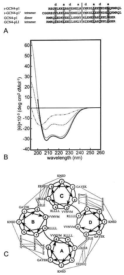
(A) Sequence alignment of the true 35-residue retro-leucine zipper based on the sequence of GCN4-p1, previously termed r-LZ35 (r-GCN4-p1), r-GCN4-p1′, wild-type C-terminal 33-residue leucine zipper moiety of the yeast transcription activator GCN4 (GCN4-p1), and GCN4-p1 mutant with leucine and isoleucine residues in positions a and d, respectively (GCN4-pLI). Because of the low sequence identity between r-GCN4-p1′ and GCN4-p1 of 19% and the symmetry of the folds, there is no unique superposition. The structures could be shifted by seven residues along the superhelix axis, yielding superpositions with similar rms deviations (rmsds). For this alignment, the conserved central cavity was taken into account. The palindrome axis and the residues that participate in the seven-residue repeats are indicated. Both GCN4-p1 and GCN4-pLI are acetylated at the N terminus. (B) CD spectra of r-GCN4-p1 at various concentrations (1 mM, dotted line; 120 μM, dashed line; 30 μM, dotted/dashed line) and oxidized r-GCN4-p1′ (15 μM, solid line). (C) Helical wheel of the r-GCN4-p1′ tetramer. Helical wheel representation of residues 4 to 36. The view is from the N terminus. Heptad positions are labeled a through g. Polar interactions between side chains are indicated by dashed lines. Main-chain side-chain polar interactions are shown by continuous lines.
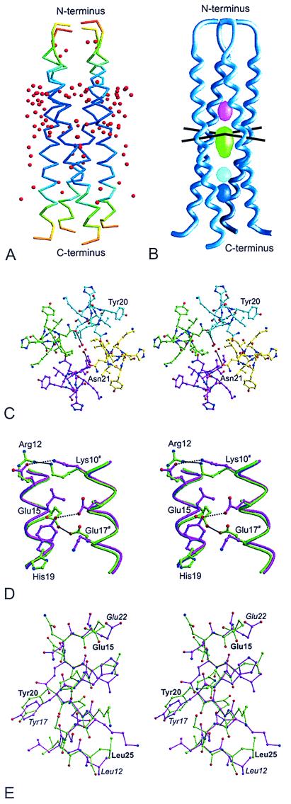
(A) Backbone of the r-GCN4-p1′ tetramer colored according to temperature factor (dark blue = 20 Å2; red = 100 Å2). Water molecules are shown as red bullets. Figures were prepared with the programs molscript and bobscript (18, 19). (B) Cavities in the r-GCN4-p1′ structure. The volumes of the cavities are 87 Å3 (green), 39 Å3 (blue), 36 Å3 (magenta), and 30 Å3 (cyan). The cavities were calculated with a probe radius of 1.6 Å. The palindrome axis intersects the 4-fold symmetry axes at the top of the large cavity; axes are indicated as black lines. The figure was generated with the program grasp (22). (C) Stereo projection of water molecules in the central cavity. The view is parallel to the 4-fold symmetry axis. Possible hydrogen bonds are indicated by dashed lines. (D) Polar interactions on the surface of r-GCN4-p1′ (green carbon atoms) and GCN4-pLI mutant (pink carbon atoms). Polar interactions are indicated by black lines. Residue numbering corresponds to the r-GCN4-p1′ structure; # refers to the next repeat. (E) Superposition of residues 15–25 of the r-GCN4-p1′ structure (green carbon atoms) onto residues 12–22 of the wild-type GCN4-p1 structure (pink carbon atoms, residue numbers in italics). The Cα atoms superimpose with an rmsd of 0.29 Å. In the r-GCN4-p1′ structure, the direction of the sequence is from top to bottom. In the GCN4-p1 structure, the direction is from bottom to top.
References
-
- Anfinsen C B. Science. 1973;181:223–230. - PubMed
-
- Guptasarma P. FEBS Lett. 1992;310:205–210. - PubMed
-
- Olszewski K A, Kolinski A, Skolnick J. Protein Eng. 1996;9:5–14. - PubMed
-
- Witte K, Skolnick J, Wong C-H. J Am Chem Soc. 1998;120:13042–13046.
-
- Beck K, Brodsky B. J Struct Biol. 1998;122:17–29. - PubMed
Publication types
MeSH terms
Substances
Associated data
- Actions
LinkOut - more resources
Full Text Sources
Other Literature Sources
Molecular Biology Databases
