. 2000 Mar 14;97(6):2626-31.
doi: 10.1073/pnas.97.6.2626.

Activin receptor-like kinase 1 modulates transforming growth factor-beta 1 signaling in the regulation of angiogenesis

Affiliations

Activin receptor-like kinase 1 modulates transforming growth factor-beta 1 signaling in the regulation of angiogenesis

S P Oh et al. Proc Natl Acad Sci U S A. .

Abstract

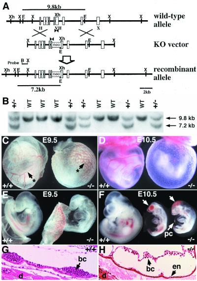
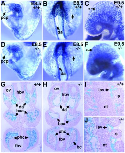
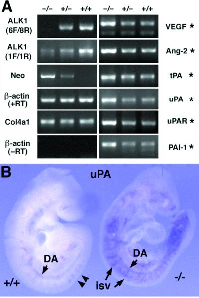
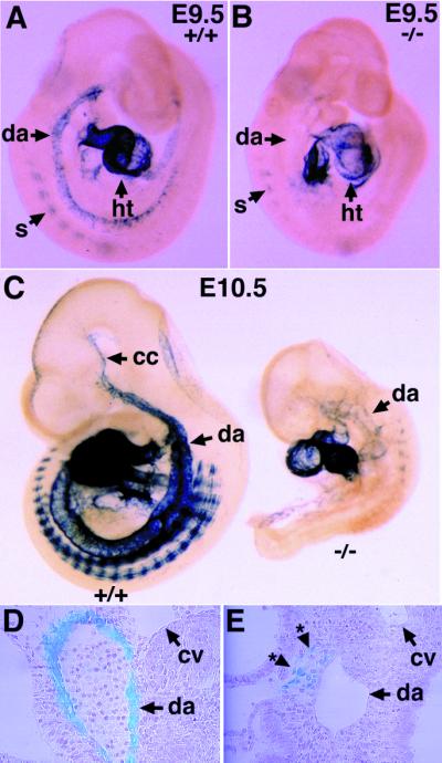
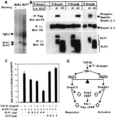
The activin receptor-like kinase 1 (ALK1) is a type I receptor for transforming growth factor-beta (TGF-beta) family proteins. Expression of ALK1 in blood vessels and mutations of the ALK1 gene in human type II hereditary hemorrhagic telangiectasia patients suggest that ALK1 may have an important role during vascular development. To define the function of ALK1 during development, we inactivated the ALK1 gene in mice by gene targeting. The ALK1 homozygous embryos die at midgestation, exhibiting severe vascular abnormalities characterized by excessive fusion of capillary plexes into cavernous vessels and hyperdilation of large vessels. These vascular defects are associated with enhanced expression of angiogenic factors and proteases and are characterized by deficient differentiation and recruitment of vascular smooth muscle cells. The blood vessel defects in ALK1-deficient mice are reminiscent of mice lacking TGF-beta1, TGF-beta type II receptor (TbetaR-II), or endoglin, suggesting that ALK1 may mediate TGF-beta1 signal in endothelial cells. Consistent with this hypothesis, we demonstrate that ALK1 in endothelial cells binds to TGF-beta1 and TbetaR-II. Furthermore, the ALK1 signaling pathway can inhibit TGF-beta1-dependent transcriptional activation mediated by the known TGF-beta1 type I receptor, ALK5. Taken together, our results suggest that the balance between the ALK1 and ALK5 signaling pathways in endothelial cells plays a crucial role in determining vascular endothelial properties during angiogenesis.


Figures

Figure 1
Figure 1
Targeted disruption of the mouse ALK1 gene results in embryonic lethality. (A) Schematic diagram (from top to bottom) of the wild-type allele, the knockout (KO) vector, and the recombinant mutant allele. Exons are indicated by rectangles and roman numerals. The PGK-neo cassette was inserted into the XhoI site of exon VII encoding kinase subdomain V (10). An EcoRI site was introduced into the target ALK1 locus, thus the 9.8-kb and 7.2-kb EcoRI fragments indicated by lines represent wild-type and mutant alleles, respectively. PCR primers (arrowheads) and the 5′ external probe (B-X) for Southern hybridization are indicated. B, BamHI; E, EcoRI; S, SalI; X, XbaI; Xh, XhoI. (B) Southern blot analysis of tail DNA of a litter of newborns from the ALK1+/− intercross. (C-F) Gross morphology of yolk sac (C, D) and embryo proper (E, F) of wild-type (Left) and mutant embryos (Right) at E9.5 (C, E) and E10.5 (D, F). Arrows with asterisks (C) indicate the yolk sac blood vessels. Arrows (F) indicate clumps of blood cells in the mutant embryos. pc, pericardium. (G, H) Histological analysis of E9.5 wild type (G) and mutant (H) embryos in decidua. In the mutant yolk sac, blood, and ECs are present, but the vessels are enlarged (H). bc, blood cells; d, decidua; en, endothelium.
Figure 2
Figure 2
Dilation of major vessels and fusion of capillaries in ALK1−/− embryos. Developing blood vessels of [ALK1+/+, Flk1+/−] (A–C, G, I) and [ALK1−/−, Flk1+/−] (D–F, H, J) embryos are visualized by X-Gal staining (blue). (A, B, D, and E) Lateral (A) and posterior (B) views of a normal E8.5 embryo and lateral (D) and posterior (E) views of an ALK1−/− littermate. Note the vascular patterns, in particular the primary capillary plexus (pcp) in head and the yolk sac indicated by arrows, are essentially indistinguishable between normal and mutant embryos. (C and F) Gross vascular morphology of the head region in E9.5 embryos. Note the absence of a capillary network in the mutant embryo indicated by an arrow with an asterisk (F). (G–J) Histological sections of E9.5 [ALK1+/+, Flk1+/−] (G, I) and [ALK1−/−, Flk1+/−] (H, J) embryos after whole-mount X-Gal staining followed by plastic embedding. (G) Transverse sections of a normal embryo at otic vesicle (ov) region show Flk+ cells in the endothelium of the dorsal aorta (da), branchial arch arteries (baa), and peripheral head capillaries (phc). (H) Transverse sections of a mutant embryo in corresponding to G. Note that dorsal aorta and branchial arch arteries were greatly dilated and fused with surrounding capillaries, forming large cavernous vessels. The endothelial linings of the mutant blood vessels were intact, suggesting that the clumps of blood cells seen in the mutant embryos (Fig. 1F) result not from hemorrhages but probably from impaired circulation of blood in malformed vessels. (I) A normal embryo section showing neural tube (nt), somites (s), and intersomitic vessels (isv). (J) Sections of mutant embryos corresponding to I. Note the dilation and fusion of intersomitic vessels in the mutant embryos. a, allantois; bc, blood cells; fbv, forebrain vesicle; hbv, hind brain vesicle.
Figure 3
Figure 3
Elevated expression of angiogenic factors and plasminogen activators in the ALK1−/− embryos. (A) Visualized amplified PCR products by the ethidium bromide staining. The same amount of cDNA and methods were used for PCR amplification of each gene. After 40 cycles of amplification and quantitative analyses, PCR products were separated on an agarose gel. All the ethidium bromide staining patterns reflecting amplification plots as represented in supplemental Fig. 6 (www.pnas.org) are shown. ALK1 primers (6F/8R) spanning the neor insertion site did not amplify a DNA band from −/− embryos. ALK1 primers (1F/1R) upstream of the neor insertion site amplified a DNA band from −/− embryos in a greatly diminished level, indicating that a reduced level of transcripts is made in the mutant allele. Conversely, neo gene primers amplified cDNA from −/− and +/− embryos but not from the wild-type embryos. Note that the intensity of the amplified signal for ALK1 and Neo was gene-dosage dependent. The transcript level of the α1 type IV collagen (Col4a1) gene was unaltered, regardless of the genotypes of embryos, whereas, transcript levels of VEGF, Ang-2, tissue-type PA, uPA, uPA-receptor, and PAI-1 were significantly elevated in the ALK1−/− embryos (indicated by asterisks). (B) Whole-mount in situ hybridization of the uPA gene in the E9.5 wild-type (Left) and ALK1−/− (Right) embryos. uPA was detected in the branching points (arrow heads) of the dorsal aorta and ISV of wild-type embryo, whereas overexpressed uPA expression was detected in the various developing blood vessels in the mutant embryos.
Figure 4
Figure 4
Delayed differentiation and improper localization of vascular smooth muscle cells in the ALK1−/− embryos. (A and B) Expression of tg-SM in E9.5 wild-type (A) and ALK1−/− mutant (B) embryos. Note the absence of tg-SM expression in the dorsal aorta (arrow with asterisk) of the mutant embryos (B). (C) Expression of the tg-SM in E10.5 wild-type (Left) and ALK1−/− mutant (Right) embryos. The mutant embryo is severely growth retarded and expresses tg-SM in the anterior part of the dorsal aorta and somites. (D and E) Plastic sections of embryos after X-Gal staining shown in C. Note the dorsal aorta in the wild-type embryo is surrounded by lacZ-positive vascular smooth muscle cells (D), whereas the lacZ-positive cells (*) in the mutant embryo are localized near the dorsal aorta but fail to encircle the vessel (E). cc, common carotid; cv, cardinal vein; da, dorsal aorta; ht, heart; s, somite.
Figure 5
Figure 5
ALK1 binds to TGF-β1 in ECs, activates Smad1 and Smad5, and can inhibit the TGF-β1-responsive 3TP-Lux induction. (A) TGF-β1 binds to ALK1 as well as ALK5 (TβR-I) in HUVEC. HUVEC were affinity crosslinked with 125I-TGF-β1 and immunoprecipitated by using antibodies specific to either ALK1 or ALK5. Blocking of ALK1 antiserum was done in the presence of the peptide used for immunization of the rabbit (15). The ALK1 band migrated slightly slower than ALK5, in agreement with the difference in the sizes of ALK1 and ALK5. (B) ALK1 phosphorylates Smad1 and Smad5 but not Smad2. COS cells were transfected with Flag-tagged (F) Smad1, Smad5, or Smad2, and constitutively active (c.a.) forms of ALK1 (A1) or ALK5 (A5). (Top). Phosphorylation of Smads was detected by immunoprecipitation of each Smad with a Flag antibody followed by immunoblotting by using an antiphosphoserine antiserum. (Center and Bottom). Expression of Smads and ALKs was determined by anti-Flag and antihemagglutinin antibodies, respectively. (C) ALK1 inhibits TGF-β1-dependent 3TP-Lux induction mediated by ALK5 in HepG2 cells. A TGF-β responsive element linked to the luciferase gene (3TP-Lux) was transfected into HepG2 cells together with increasing amounts of ALK1 or ALK2 in the presence of ALK5. After transfection, cells were treated with (solid bars) or without (open bars) TGF-β1 (5 ng/ml) for 24 hr. Luciferase activities were then measured and plotted. In all assays, luciferase activities are plotted in arbitrary units. (D) The balance model for TGF-β1 signaling in regulation of angiogenesis. Explanation is in the text.

References

    1. Hoodless P A, Wrana J L. Curr Top Microbial Immunol. 1998;228:235–272. - PubMed
    1. Risau W. Nature (London) 1997;386:671–674. - PubMed
    1. Pepper M S. Cytokines Growth Factor Rev. 1997;8:21–43. - PubMed
    1. Gajdusek C M, Luo Z, Mayberg M R. J Cell Physiol. 1993;157:133–144. - PubMed
    1. Pepper M S, Vassalli J-D, Orci L, Montesano R. Exp Cell Res. 1993;204:356–363. - PubMed

Publication types

MeSH terms