. 2000 Mar 15;523 Pt 3(Pt 3):741-53.
doi: 10.1111/j.1469-7793.2000.00741.x.

Non-linear membrane properties of sacral sphincter motoneurones in the decerebrate cat

Affiliations

Non-linear membrane properties of sacral sphincter motoneurones in the decerebrate cat

K L Paroschy et al. J Physiol. .

Abstract

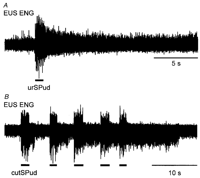
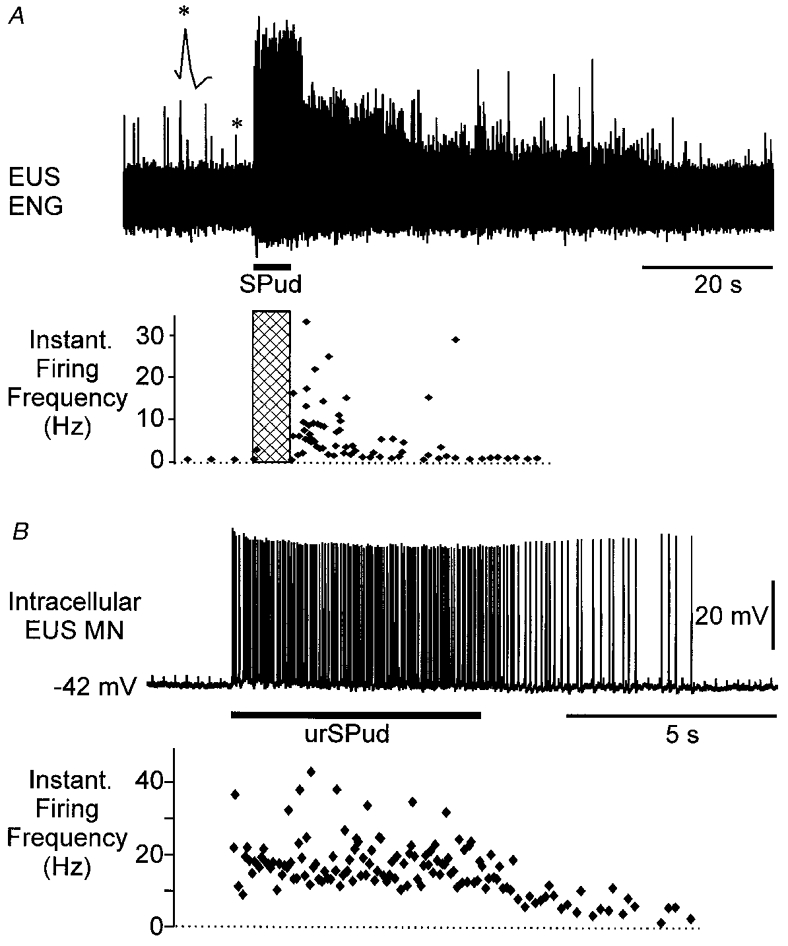
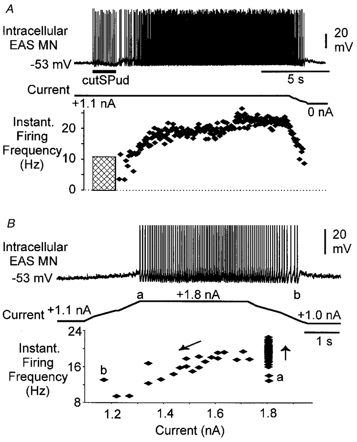
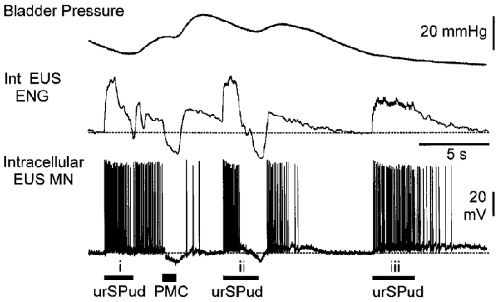
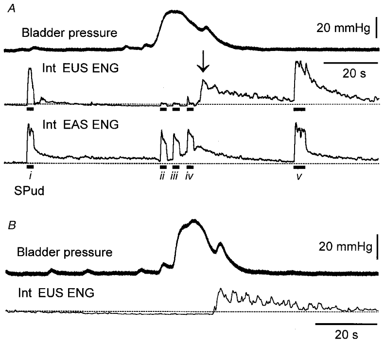
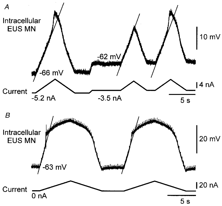
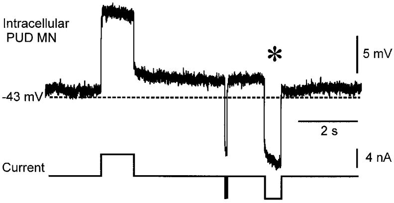
1. Responses to pudendal afferent stimulation and depolarizing intracellular current injection were examined in sacral sphincter motoneurones in decerebrate cats. 2. In 16 animals examined, 2-10 s trains of electrical stimulation of pudendal afferents evoked sustained sphincter motoneurone activity lasting from 5 to >50 s after stimulation. The sustained response was observed in: 11 animals in the absence of any drugs; two animals after the intravenous administration of 5-hydroxytryptophan (5-HTP; <= 20 mg kg-1); one animal in which methoxamine was perfused onto the ventral surface of the exposed spinal cord; and two animals following the administration of intravenous noradrenergic agonists. 3. Extracellular and intracellular recordings from sphincter motoneurones revealed that the persistent firing evoked by afferent stimulation could be terminated by motoneurone membrane hyperpolarization during micturition or by intracellular current injection. 4. Intracellular recordings revealed that 22/40 sphincter motoneurones examined displayed a non-linear, steep increase in the membrane potential in response to depolarizing ramp current injection. The mean voltage threshold for this non-linear membrane response was -43 +/- 3 mV. Five of the 22 cells displaying the non-linear membrane response were recorded prior to the administration of 5-HTP; 17 after the intravenous administration of 5-HTP (<= 20 mg kg-1). 5. It is concluded that sphincter motoneurones have a voltage-sensitive, non-linear membrane response to depolarization that could contribute to sustained sphincter motoneurone firing during continence.


Figures

Figure 1
Figure 1. Stimulus-locked and sustained EUS ENG activity evoked by stimulation of pudendal afferents
A, a 10 Hz train of stimuli (5T) to the urSPud nerve evoked multi-unit ENG activity during the period of stimulation (indicated by bar below the ENG) that persisted for seconds after the stimulation. B, response in another animal in which repeated trains (10 Hz, 5T) of stimulation of the cutSPud nerve evoked sustained responses that increased in duration with subsequent stimulation. Neither animal had been given 5-HTP, methoxamine or noradrenaline.
Figure 2
Figure 2. Sustained responses and increased firing frequency in EUS units following afferent stimulation
A, a single unit (*, enlarged above trace) discriminated from the population ENG (upper panel) and the instantaneous firing frequency of the unit following stimulation of the SPud nerve at 10 Hz (5T, hatched bar) (lower panel). No drugs had been given to this animal. B, intracellular recording from an EUS motoneurone (MN, upper panel, resting membrane potential (Vm) -42 mV) revealed that this cell fired at ≈20 Hz during stimulation of the urSPud nerve (5T, 4 Hz) and that the firing continued at ≈8 Hz for almost 5 s after stimulation. In this animal, intravenous noradrenaline had been given to elevate the blood pressure.
Figure 3
Figure 3. Effect of membrane potential on afferent-evoked persistent firing in an EAS motoneurone
A, intracellular EAS motoneurone recording (upper panel, resting Vm -57 mV) shows that when the cell was depolarized to -53 mV with 1.1 nA of bias current, persistent firing was evoked by cutSPud afferent stimulation (10T, 10 Hz). A plot of the instantaneous firing frequency of the motoneurone (lower panel) revealed the persistent firing (≈18 Hz) lasted several seconds then increased to 21 Hz. Reducing the depolarizing bias current resulted in a drop off in the persistent firing of the cell. The hatched region represents the period of stimulation when doublet and triplet firing was evoked by each stimulus but was not included in this plot of instantaneous firing frequency. B, in this same cell, current injection alone produced persistent 18 Hz firing that terminated when the membrane was hyperpolarized (i.e. depolarizing bias current removed). Spikes have been truncated for illustration purposes. The plot of the instantaneous firing versus current (lower panel) shows that firing started at 1.8 nA of current (a), accelerated (upward arrow) then persisted as the current was decreased (downward arrow) to below (b) the initial current (a) that activated the cell. This animal had been given 16 mg kg−1 5-HTP 2.5 h prior to the recording.
Figure 6
Figure 6. Effects of synaptically evoked membrane hyperpolarization on persistent motoneurone firing
Bladder pressure, integrated (Int) EUS ENG and intracellular EUS motoneurone records (resting Vm -58 mV) during stimulation of the urSPud nerve and pontine micturition centre (PMC) are shown. Trains of stimuli to the urSPud nerve (i; 10T, 20 Hz) evoked EUS firing that persisted until the motoneurone membrane was hyperpolarized by PMC stimulation (50 Hz, 200 μA). The second train of urethral stimulation (ii) evoked stimulus-locked firing that stopped when the membrane hyperpolarized and resumed after the stimulation and hyperpolarization was terminated. The final train (iii) of urSPud stimulation (10T, 10 Hz) evoked firing that persisted for several seconds before stopping spontaneously. The dashed line indicates the level of ENG activity and the motoneurone membrane potential prior to any stimulation. This animal had received 8.4 mg kg−1 5-HTP 30 min before the recording.
Figure 7
Figure 7. Suppression of afferent-evoked sustained efferent responses during distension-evoked micturition
A, distension of the bladder evoked a micturition reflex characterized by an increase in bladder pressure (upper trace) during which time there was a suppression of EUS ENG activity (second trace, 1 Hz integrated) that was not seen in the EAS ENG activity (third trace). The dashed line indicates the pre-void level of ENG activity. SPud nerve stimulation (5T, 10 Hz; indicated by bar below trace) evoked sustained EUS activity prior to (i) and after (v) the void. During micturition (ii-iv), both stimulus-locked and sustained EUS ENG responses evoked by afferent stimulation were attenuated while the evoked EAS ENG responses were largely unchanged. As the bladder contraction ended, an increase in EUS ENG activity (arrow) was evident. B, in this same animal, the post-void increase in EUS activity was also evident in the absence of peripheral nerve stimulation.
Figure 4
Figure 4. Non-linear changes in membrane potential in response to current injection
A, changes in membrane potential (upper trace) in response to ramp current injections of varying sizes (lower trace) are shown in EUS motoneurones recorded with QX-314 in the micropipette. Depolarizing current sufficient to bring the Vm to approximately -47 mV resulted in an abrupt change in the slope of the membrane trajectory (indicated by continuous diagonal line). The middle current ramp was not of sufficient size to evoke this non-linear response. This animal had been given 4 mg kg−1 5-HTP prior to the recording. B, another EUS motoneurone displayed a non-linear membrane response with longer ramp injections; no 5-HTP had been administered in this animal. In A, the motoneurone resting Vm was -55 mV; in B it was -61 mV.
Figure 5
Figure 5. Persistent membrane depolarization in response to depolarizing square wave pulses
In this pudendal motoneurone (resting Vm -43 mV), depolarizing pulses produced a persistent membrane depolarization of several millivolts. The depolarization was terminated by a hyperpolarizing current injection longer than 200 ms (*) but not by a very brief pulse of the same amplitude. QX-314 (50 mM) was included in the recording micropipette solution. This animal had received 8 mg kg−1 5-HTP ≈40 min prior to the recording.

References

    1. Beattie MS, Li Q, Leedy MG, Bresnahan JC. Motoneurons innervating the external anal and urethral sphincter of the female cat have different patterns of dendritic arborisation. Neuroscience Letters. 1990;111:69–74. - PubMed
    1. Bennett DJ, Hultborn H, Fedirchuk B, Gorassini M. Synaptic activation of plateaus in hind limb motoneurones of decerebrate cats. Journal of Neurophysiology. 1998a;80:2023–2037. - PubMed
    1. Bennett DJ, Hultborn H, Fedirchuk B, Gorassini M. Short-term plasticity in hind limb motoneurones of decerebrate cat. Journal of Neurophysiology. 1998b;80:2038–2045. - PubMed
    1. Binder MD, Heckman CJ, Powers RK. The physiological control of motoneuron activity. In: Rowell LB, Shepherd JT, Smith J, editors. Handbook of Physiology, Neural Control of Movement. Vol. 1. Oxford: Oxford University Press; 1996. pp. 3–53. assoc.
    1. Bishop B, Garry RC, Roberts TDM, Todd JK. Control of the external sphincter of the anus of the cat. The Journal of Physiology. 1956;134:229–240. - PMC - PubMed

Publication types

MeSH terms

Substances

LinkOut - more resources