. 2000 Mar 15;523 Pt 3(Pt 3):771-83.
doi: 10.1111/j.1469-7793.2000.t01-1-00771.x.

Surround inhibition of mammalian AII amacrine cells is generated in the proximal retina

Affiliations

Surround inhibition of mammalian AII amacrine cells is generated in the proximal retina

S A Bloomfield et al. J Physiol. .

Abstract

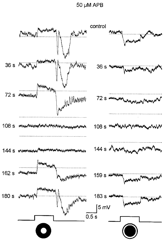
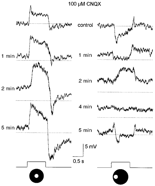
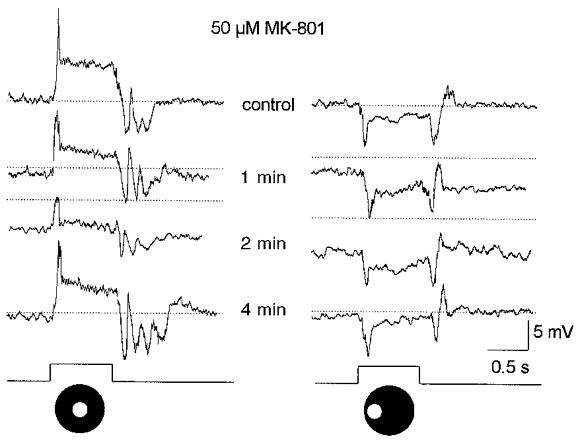
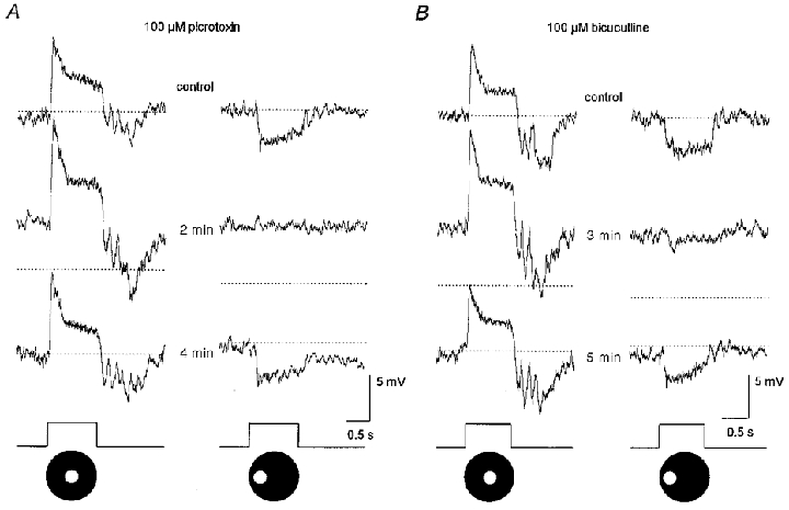
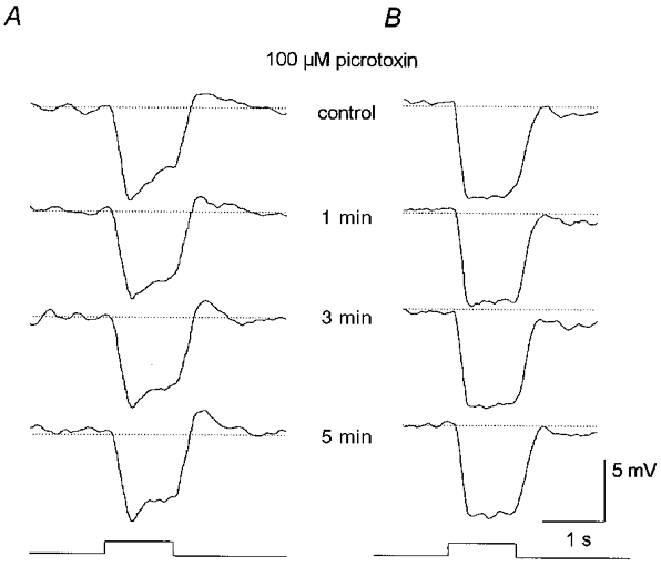
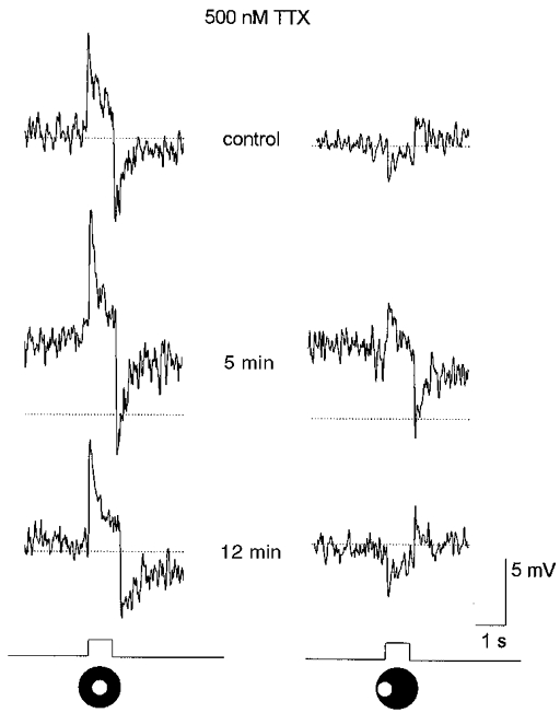
1. Intracellular recordings were obtained from neurons in the superfused retina-eyecup preparation of the rabbit under dark-adapted conditions. Neurotransmitter agonists and antagonists were applied exogenously via the superfusate to dissect the synaptic pathways pharmacologically and thereby determine those pathways responsible for the generation of the on-centre/off-surround receptive fields of AII amacrine cells. 2. Application of the metabotropic glutamate receptor agonist, APB, reversibly blocked both the on-centre and off-surround responses of AII cells. These data were consistent with the idea that both the centre- and surround-mediated responses are derived from inputs from the presynaptic rod bipolar cells. 3. Whereas rod bipolar cells showed on-receptive fields approximately 100 microm across, we found no evidence for an antagonistic off-surround response using light stimuli which effectively elicited the off-surrounds of AII amacrine cells. These results indicated that the surrounds of AII cells are not derived from rod bipolar cell inputs. 4. Application of the ionotropic glutamate receptor antagonists CNQX or DNQX enhanced the on-centre responses of AII cells but attenuated the off-surround responses. These data indicated that the centre- and surround-mediated responses could not both be derived from signals crossing the rod bipolar-to-AII cell synapse. 5. Application of the glycine antagonist, strychnine, had only minor and variable effects on AII cell responses. However, the GABA antagonists picrotoxin and bicuculline enhanced the on-centre response but attenuated or completely blocked the off-surround response of AII cells. The GABA antagonists had no effect on the responses of horizontal cells indicating that their effects on AII cell responses reflected actions on inner retinal circuitry rather than feedback circuitry in the outer plexiform layer. 6. Application of the voltage-gated sodium channel blocker TTX enhanced the on-centre responses of AII cells but attenuated or abolished their off-surround responses. 7. Taken together, our results suggest that the on-centre responses of AII cells result from the major excitatory drive from rod bipolar cells. However, the surround receptive fields of AII cells appear to be generated by lateral, inhibitory signals derived from neighbouring GABAergic, on-centre amacrine cells. A model is presented whereby the S1 amacrine cells produce the surround receptive fields of AII amacrine cells via inhibitory, feedback circuitry to the axon terminals of rod bipolar cells.
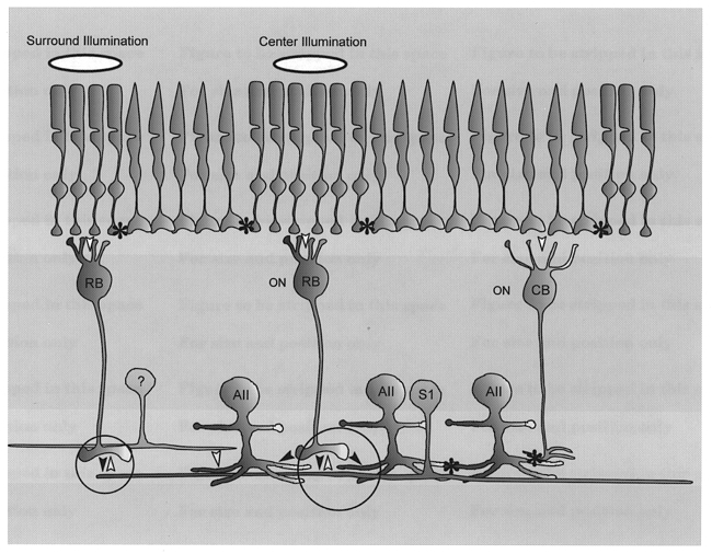

Figures

Figure 1
Figure 1. Light-evoked responses of AII amacrine cells in the dark-adapted retina
A, typical response consists of a transient depolarization at light onset followed by a sustained phase and a large-amplitude, oscillating hyperpolarization at light offset. Stimulus is a 75 μm diameter spot of light. Stimulus intensity, log-5.5; maximum intensity (log0.0 = 2.37 mW cm−2). Trace at bottom indicates onset and offset of the light stimulus. B, on occasion spike-like waves could be seen in the responses of AII amacrine cells. These spikes could be abolished with TTX indicating that they are generated by voltage-gated sodium channels. Stimulus parameters are the same as in A.
Figure 2
Figure 2. Receptive field properties of dark-adapted AII amacrine cells
A, responses to a 50 μm wide, 6.0 mm long rectangular slit of light which was moved in discrete steps across the retinal surface. At 0 μm the slit was centred over the cell. The values to the left of each trace represent how far off-centre the slit was positioned; polarity of number indicates direction of movement. This cell showed an on-centre receptive field measured at 66 μm across. Stimulus intensity, log-5.5. Trace at bottom indicates presentation of light stimulus. B, on-centre response of an AII cell to a centred 75 μm diameter spot of light. Stimulus intensity, log-5.5. C, off-surround response of same AII cell as in B after the spot stimulus was displaced laterally by 100 μm.
Figure 3
Figure 3. Effects of 50 μM APB on the responses of a dark-adapted AII cell
Prior to drug application (control), the cell responded to the 75 μm diameter spot of light (left panel) with the typical on-centre response components. Presentation of an annular light stimulus (inner diameter, 75 μm; outer diameter, 350 μm) (right panel) evoked a clear off-surround response. The values to the left of each trace indicate time from the beginning of the 40 s long application of APB; for example, the response at 72 s corresponds to 32 s after return to the control superfusate. APB produced an approximate 10 mV hyperpolarization and reversibly blocked both the on-centre and off-surround responses. Dotted lines correspond to the dark membrane potential during the initial control response and are provided for comparison. Intensity of both spot and annular stimuli, log-5.5. Traces at bottom indicate presentation of light stimuli.
Figure 4
Figure 4. Responses of a rod bipolar cell to different stimulus configurations
A, responses to a 50 μm wide, 6.0 mm long rectangular slit of light moved in discrete steps across the retinal surface. Conventions are the same as in Fig. 2A. The on-centre receptive field was measured to be ≈100 μm across. Stimulus intensity, log-5.5. B, responses to a 75 μm diameter spot of light and an annulus of light (inner diameter, 75 μm; outer diameter, 350 μm). Conventions are the same as in Fig. 3. Both stimuli produced an on-centre response with the annulus failing to produce an off-surround. Stimulus intensity, log-5.5. C, responses to a 75 μm diameter spot of light first centred over the cell and then displaced laterally by 100 μm. Conventions are the same as in Fig. 2B and C. In both cases the spot evoked an on-centre response with the displaced stimulus failing to produce an off-surround response. Stimulus intensity, log-5.5. Taken together, these data indicate that rod bipolar cells do not express off-surround receptive fields.
Figure 5
Figure 5. Effects of 100 μM CNQX on AII cell responses
Prior to application of CNQX, a 75 μm diameter spot of light evoked an on-centre response (left panel), whereas the same spot displaced laterally by 100 μm evoked an off-surround response (right panel). Conventions are the same as in Fig. 3. A 40 s application of CNQX produced an approximate 5 mV depolarization and reversibly enhanced the on-centre response to the centred spot of light. CNQX abolished the off-surround response to the displaced spot of light, replacing the hyperpolarizing response with a depolarization. The depolarizing response is believed to be an on-centre response, evoked by light scatter into the centre receptive field, that was revealed after abolition of the off-surround. Note that the drug effects on the off-surround responses were reversed prior to that of the on-centre response. Stimulus intensity, log-5.5. Traces at bottom indicate presentation of the light stimuli.
Figure 6
Figure 6. Effects of 50 μM MK-801 on AII cell responses
A 40 s application of MK-801 produced an approximate 5 mV hyperpolarization of the membrane and reduced the on-centre response by ≈60 % (left panel) but had no obvious effect on the off-surround response. Stimuli consisted of a 75 μm diameter spot of light centred over the cell (left panel) and then translated laterally by 100 μm (right panel). Stimuli intensity, log-5.5. Drug effects were reversed within approximately 3 min after return to the control superfusate. Conventions are the same as in Fig. 3. Traces at bottom indicate presentation of the light stimuli.
Figure 7
Figure 7. Effects of GABA antagonists on AII cell responses
A, a 40 s application of 100 μm picrotoxin produced a 5 mV depolarization of the membrane, enhanced the on-centre response of the AII cell but completely abolished the off-surround response. These drug effects were reversed approximately 3 min after return to the control superfusate. Stimuli consisted of a 75 μm diameter spot of light centred over the cell (left panel) and displaced laterally by 100 μm (right panel). Conventions are the same as in Fig. 3. B, a 40 s application of 100 μM bicuculline also depolarized the membrane by ≈5 mV and enhanced the on-centre response. However, in contrast to the effects of picrotoxin, bicuculline markedly reduced but did not abolish the off-surround response. Stimulus parameters are the same as in A. Traces at bottom indicate presentation of light stimuli.
Figure 8
Figure 8. Effects of 100 μM picrotoxin on B-type horizontal cell responses
A, effects of a 2 min application of picrotoxin on the responses recorded from the somatic ending of a B-type horizontal cell. The drug showed no effect on either the membrane potential or light-evoked responses. Stimulus was full-field illumination with intensity of log-5.5. Conventions are the same as in Fig. 3. B, effects of a 2 min application of picrotoxin on the responses recorded from the axon terminal ending of a B-type horizontal cell. Again, the drug had no clear effects on this cell. Stimulus parameters are the same as in A. Traces at bottom indicate presentation of light stimuli.
Figure 9
Figure 9. Effects of 500 nM TTX on AII cell responses
A 3 min application of TTX produced a 5 mV depolarization of the membrane and enhanced the on-centre response. TTX also reduced the off-surround response, replacing it with a depolarization with waveform similar to that of the on-centre response, albeit smaller in amplitude. Analogous to the effects of CNQX, we believe that the depolarizing response is an on-centre response, evoked by light scatter into the centre receptive field, that was revealed after reduction or abolition of the off-surround. Stimuli are a 75 μm diameter spot of light centred over the cell (left panel) and displaced laterally by 100 μm (right panel). Conventions are the same as in Fig. 3. Traces at bottom indicate presentation of light stimuli.
Figure 10
Figure 10. Schematic diagram of synaptic circuitry subserving AII cell centre and surround responses in the dark-adapted retina
Illumination of the centre receptive field of an AII cell produces an on-centre responses via the rod → rod bipolar cell → AII amacrine cell pathway (large circle). Illumination of the surround receptive field produces an off-surround response by the pathway rod → rod bipolar cell → S1 amacrine cell (small circle) which carries the signal centrally and feeds back via a GABAergic inhibitory (sign inverting) synapse to central rod bipolar cell axon terminals (large circle). Activation of this ‘surround-generating’ circuit shunts the signal in the rod bipolar cell axon terminal thereby reducing the glutamate release onto AII cells and producing the hyperpolarizing off-response. An alternative, but less likely, pathway for surround inhibition may be from unidentified amacrine cells (?) which provide direct inhibitory inputs onto AII amacrine cell dendrites. Asterisks, gap junctions; filled arrowheads, sign-conserving synapse; open arrowheads, sign-inverting synapse; AII, AII amacrine cell; RB, rod bipolar cell; S1, S1 amacrine cell; ON, on-centre.

References

    1. Barlow HB, Fitzhugh R, Kuffler SW. Change of organization of receptive fields of the cat's retina during dark adaptation. The Journal of Physiology. 1957;137:338–354. - PMC - PubMed
    1. Baylor DA, Fuortes MGF, O'Bryan PM. Receptive field of cones in the retina of the turtle. The Journal of Physiology. 1971;214:265–294. - PMC - PubMed
    1. Bloomfield SA. Relationship between receptive and dendritic field size of amacrine cells in the rabbit retina. Journal of Neurophysiology. 1992;68:711–725. - PubMed
    1. Bloomfield SA. Effect of spike blockade on the receptive-field size of amacrine and ganglion cells in the rabbit retina. Journal of Neurophysiology. 1996;75:1878–1893. - PubMed
    1. Bloomfield SA, Dowling JE. Roles of aspartate and glutamate in synaptic transmission in rabbit retina. I. Outer plexiform layer. Journal of Neurophysiology. 1985a;53:699–713. - PubMed

Publication types

MeSH terms

LinkOut - more resources