. 2000 Feb;81(1):51-6.
doi: 10.1046/j.1365-2613.2000.00140.x.

Warfarin exposure and calcification of the arterial system in the rat

Affiliations

Warfarin exposure and calcification of the arterial system in the rat

A M Howe et al. Int J Exp Pathol. 2000 Feb.

Abstract

There is evidence from knock-out mice that the extrahepatic vitamin K-dependent protein, matrix gla protein, is necessary to prevent arterial calcification. The aim of this study was to determine if a warfarin treatment regimen in rats, designed to cause extra-hepatic vitamin K deficiency, would also cause arterial calcification. Sprague-Dawley rats were treated from birth for 5-12 weeks with daily doses of warfarin and concurrent vitamin K1. This treatment causes an extrahepatic vitamin K deficiency without affecting the vitamin K-dependent blood clotting factors. At the end of treatment the rats were killed and the vascular system was examined for evidence of calcification. All treated animals showed extensive arterial calcification. The cerebral arteries and the veins and capillaries did not appear to be affected. It is likely that humans on long-term warfarin treatment have extrahepatic vitamin K deficiency and hence they are potentially at increased risk of developing arterial calcification.


Figures
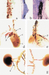
Figure 1
Figure 1
This figure shows blood vessels from rats treated with warfarin and vitamin K from birth. (b) and (c) are sections stained for calcium with von Kossa's stain and (a) and (d–g) are from whole mounts stained for calcium with alizarin red. a. Common iliac artery from a rat treated for 12 weeks from birth and killed at 1 year. The red staining corresponds to calcified areas with some circumferential arrangement. b. A section through the wall of the aorta at the level of the aortic valves (v) from a rat treated for 12 weeks. The two black areas are calcification in the aortic wall. The valve does not show any calcification. c. An enlargement of the lower area of calcification seen in 1b. d. Heart and great vessels from a rat treated for 12 weeks from birth. There is heavy staining in the pulmonary arteries (p) and coronary artery (c). The aorta (a) and its branches show less calcification. e. Heart, lungs, vessels and trachea from a rat treated for 5 weeks. The pulmonary arteries (p) extending from the lungs are heavily stained. f. Kidney and adjacent aorta and vessels from a rat treated for 12 weeks. The kidney contains a large mass of alizarin-stained material apparently in the collecting system. The material extends into the ureter (u) and appears granulated. The aorta and its branches including the renal arteries show considerable staining. g. Kidney and adjacent aorta and vessels from a rat treated for 12 weeks from birth. The aorta and its branches show considerable staining. The stained renal arteries can be seen extending into the kidney.

References

    1. Bostrum K, Watson KE, Horn S, Wortham C, Herman IM, Demer LL. Bone morphogenetic protein expressed in human atherosclerotic lesions. J. Clin. Invest. 1995;91:1800–1809. - PMC - PubMed
    1. Bucay N, Sarosi I, Dunstan CR, et al. Osteoprotegerin-deficient mice develop early onset osteoporosis and arterial calcification. Genes Devel. 1998;12:1260–1268. - PMC - PubMed
    1. Drury RAB, Wallington EA. Carleton's Histological Technique. London: Oxford University Press; 1976. pp. 150–151.
    1. Ducy P, Desbois C, Boyce B, et al. Increased bone formation in osteocalcin-deficient mice. Nature. 1996;382:448–452. - PubMed
    1. Esnouf MP, Prowse CV. The gamma-carboxy glutamic acid content of human and bovine prothrombin following warfarin treatment. Biochim. Biophys. Acta. 1977;490:471–476. - PubMed