

doi: 10.1128/AAC.44.4.1070-1074.2000.
Nucleotide sequence of the bla(RTG-2) (CARB-5) gene and phylogeny of a new group of carbenicillinases
Affiliations
- PMID: 10722515
- PMCID: PMC89816
- DOI: 10.1128/AAC.44.4.1070-1074.2000
Item in Clipboard
Nucleotide sequence of the bla(RTG-2) (CARB-5) gene and phylogeny of a new group of carbenicillinases
Antimicrob Agents Chemother.
2000 Apr.
Abstract
We determined the nucleotide sequence of the bla gene for the Acinetobacter calcoaceticus beta-lactamase previously described as CARB-5. Alignment of the deduced amino acid sequence with those of known beta-lactamases revealed that CARB-5 possesses an RTG triad in box VII, as described for the Proteus mirabilis GN79 enzyme, instead of the RSG consensus characteristic of the other carbenicillinases. Phylogenetic studies showed that these RTG enzymes constitute a new, separate group, possibly ancestors of the carbenicillinase family.
Figures
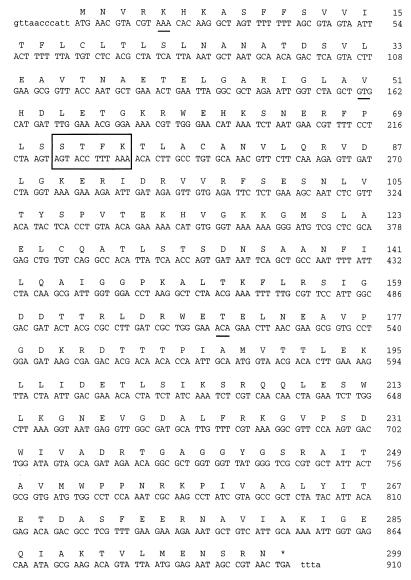
Nucleotide and deduced amino acid sequence of A. calcoaceticus β-lactamase (CARB-5). The active site is boxed, and the differences from the P. mirabilis GN79 β-lactamase sequence are underlined.
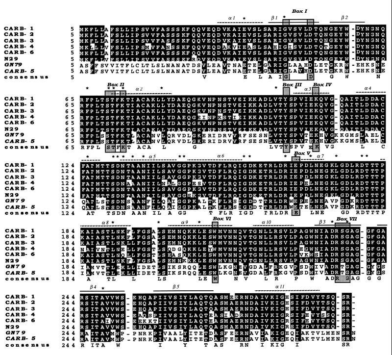
Multiple sequence alignment of the amino acid sequences of the CARB-1, CARB-2, CARB-3, CARB-4, CARB-6, P. mirabilis N29 and GN79, and CARB-5 β-lactamases. The shaded boxes (I to VII) correspond to amino acids conserved in all penicillin-recognizing enzymes, as identified by Joris et al. (15). Alpha-helix and beta-barrel motifs are from the PC1 crystal structure (3, 10). Asterisks indicate the conserved residues specific for class A β-lactamases. Amino acid changes are indicated as black letters on a white background. Sequences are numbered according to the method of Ambler (2).
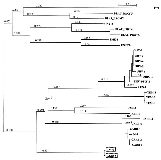
Dendrograms obtained from multiple alignment of 29 class A β-lactamases according to the neighbor-joining method (23). Branch length values represent relative phylogenetic distances.
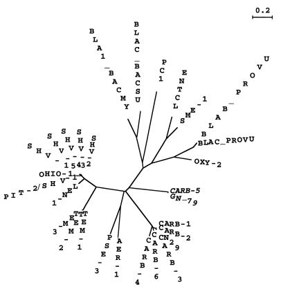
Evolutionary representation of unrooted phylogenetic tree. The phylogenetic tree was obtained as described in the legend to Fig. 3.
References
-
- Ambler R P. The structure of β-lactamases. Philos Trans R Soc Lond Biol. 1980;289:321–331. - PubMed
-
- Boissinot M, Levesque R C. Nucleotide sequence of the PSE-4 carbenicillinase gene and correlations with the Staphylococcus aureus PC1 β-lactamase crystal structure. J Biol Chem. 1990;265:1225–1230. - PubMed
MeSH terms
Substances
Associated data
- Actions
LinkOut - more resources
Full Text Sources
