. 2000 Apr;68(4):1827-33.
doi: 10.1128/IAI.68.4.1827-1833.2000.

Gamma interferon dominates the murine cytokine response to the agent of human granulocytic ehrlichiosis and helps to control the degree of early rickettsemia

Affiliations

Gamma interferon dominates the murine cytokine response to the agent of human granulocytic ehrlichiosis and helps to control the degree of early rickettsemia

M Akkoyunlu et al. Infect Immun. 2000 Apr.

Abstract

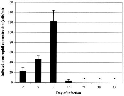
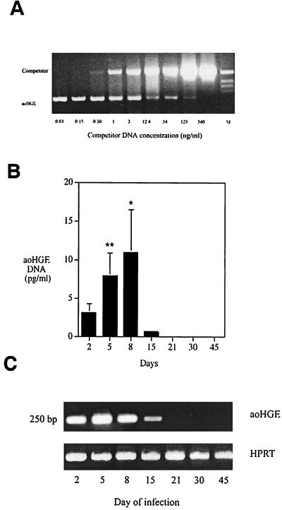
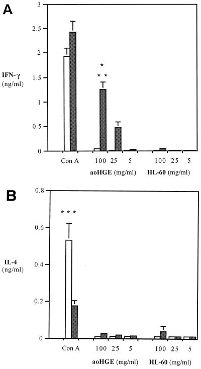
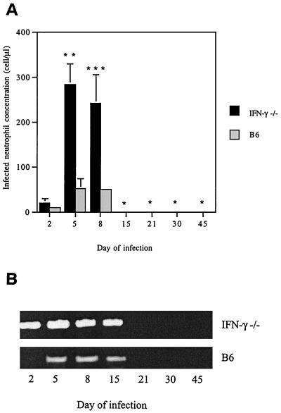
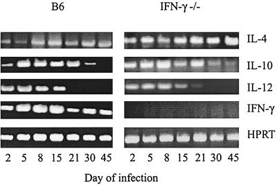
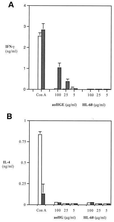
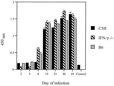
The cytokine response to the agent of human granulocytic ehrlichiosis (HGE) was assessed in a murine infection model and the role of gamma interferon (IFN-gamma), a cytokine that is crucial for host defenses against intracellular pathogens, was investigated by using IFN-gamma-deficient mice. The agent of HGE (aoHGE) is an obligate intracellular bacterium that survives within neutrophils: morulae (vacuoles containing HGE organisms) are evident in polymorphonuclear leukocytes of experimentally infected immunocompetent mice for 1 to 2 weeks. We now show that IFN-gamma levels increase during early infection of C3H/HeN or C57BL/6 mice with HGE bacteria. Moreover, in response to aoHGE extracts or concanavalin A, splenocytes from ehrlichia-infected mice produced more IFN-gamma and less interleukin-4 than controls, suggesting that aoHGE partially skewed the immune response towards a Th1 phenotype. Absolute concentration of morulae containing neutrophils in blood was 122 +/- 22 cells/microliter on day 8. The bacterial DNA burden was also highest on day 8 and then declined after IFN-gamma levels peaked. In contrast, IFN-gamma-deficient mice had a markedly elevated HGE bacteria burden with morulae concentration of 282 +/- 48 cells/microliter on day 5 (P = 0.004) and 242 +/- 63 cells/microliter on day 8 (P = 0.005). Rickettsemia resolved in immunocompetent and IFN-gamma deficient mice after 2 weeks, while both the immunocompetent and the IFN-gamma-deficient mice had increased serum antibodies against aoHGE antigens at this time point. These data demonstrate that the HGE agent elicits a prominent IFN-gamma response in mice and that IFN-gamma is important in controlling the degree of rickettsemia during the early phase of infection, while IFN-gamma independent mechanisms play a role at later time points.


Figures

FIG. 1
FIG. 1
The concentration of morula-containing neutrophils in the bloodstream of C3H mice up to 45 days after challenge. Results represent the mean ± the SD of four mice from each time point. This set of animals were infected with 100 μl of SCID mouse blood containing 195 cells of aoHGE infected neutrophils per μl. ∗, Not detected.
FIG. 2
FIG. 2
PCR analysis to determine the blood aoHGE DNA levels. (A) Representative semiquantitative competitive PCR analysis. Mouse blood DNA and a competitor DNA with primers specific for aoHGE 16S rDNA was added to the PCR reaction mixture to allow competition for the primers. Reaction tubes containing decreasing amounts of competitor DNA facilitated comparison of sample DNA and the competitor DNA. When the ratio of these two DNA bands was 1:1, they were considered equivalent, and the sample DNA concentration was equal to the competitor concentration. (B) DNA levels at different time points after infection with semiquantitative competitive PCR. Starting from day 2, aoHGE DNA concentrations increased until day 8. On day 15, aoHGE DNA levels were significantly decreased. ∗, difference in values of day 8 and day 5 are statistically insignificant (P = 0.14); ∗∗, difference in values of day 5 and day 2 are statistically significant (P = 0.001). (C) PCR analysis of blood DNA samples pooled among groups from different time points that were simultaneously amplified following an initial DNA normalization step with HPRT-based PCR.
FIG. 3
FIG. 3
(A) Splenic cytokine mRNA levels in C3H mice during infection with aoHGE. cDNA levels of each sample were normalized by using HPRT primers. (B) Serum IFN-γ concentrations measured in sandwich ELISAs. Sera of animals from each time point were pooled and applied to wells as undiluted. Means of triplicate samples are recorded. ∗, P = 0.02 compared to uninfected mice; ∗∗, P = 0.04 compared to day 2 values.
FIG. 4
FIG. 4
Levels of IFN-γ (A) and IL-4 (B) in stimulation assays with splenocytes from aoHGE-infected C3H mice (8 days). Splenocytes were incubated with 5 μg of ConA per ml and 100, 25, or 5 μg of sonication supernatants of aoHGE per ml containing HL-60 cells or HL-60 cells alone. IFN-γ levels were measured at 48 h, and IL-4 levels were measured at 24 h. All values are the means ± the SD of four mice. ∗, P < 0.002 compared to uninfected mice; ∗∗, P = 0.001 compared to HL-60 extract stimulated splenocytes; ∗∗∗, P < 0.01 compared to infected mice. □, Uninfected mice; ■, infected mice.
FIG. 5
FIG. 5
aoHGE infection in IFN-γ−/− and B/6 mice up to 45 days after challenge with ehrlichiae. (A) Morulae containing neutrophil concentrations in the bloodstream. Means ± the SD are derived from three mice examined at each time point. This set of animals were infected with 100 μl of SCID mice blood containing 198.4 cells/μl of aoHGE infected neutrophils. ∗, Not detected; ∗∗, P = 0.004 compared to B6 mice; ∗∗∗, P = 0.005 compared to B6 mice. (B) Blood aoHGE DNA levels, amplified by using 16S DNA primers. DNA levels of each sample were normalized by using HPRT (not shown).
FIG. 6
FIG. 6
Spleen cytokine mRNA levels of aoHGE-infected IFN-γ−/− and B6 mice after various intervals after aoHGE challenge. cDNA levels of each sample were normalized by using HPRT primers.
FIG. 7
FIG. 7
Levels of IFN-γ (A) and IL-4 (B) in stimulation assays with splenocytes from aoHGE-infected B6 mice (8 days). Splenocytes were incubated with 5 μg of ConA per ml and 100 μg of sonication supernatants of aoHGE per ml containing HL-60 cells or HL-60 cells alone. IFN-γ levels were measured at 48 h, and IL-4 levels were measured at 24 h. All values are the means ± the SD of four mice. □, Uninfected mice; ■, infected mice.
FIG. 8
FIG. 8
Development of anti-aoHGE IgG antibodies in the sera of C3H, IFN-γ−/−, and B6 mice. Sera were pooled among four animals from each time point. All values are the means ± the SD of three experiments.

Similar articles

Cited by

References

    1. Abbas A, Murphy K M, Sher A. Functional diversity of helper T lymphocytes. Nature. 1996;383:787–793. - PubMed
    1. Adachi J A, Grimm E M, Johnson P, Uthman M, Kaplan B, Rakita R M. Human granulocytic ehrlichiosis in a renal transplant patient: case report and review of the literature. Transplantation. 1997;64:1139–1142. - PubMed
    1. Aguero-Rosenfeld M E, Horowitz H W, Wormser G P, McKenna D F, Nowakowski J, Munoz J, Dumler J S. Human granulocytic ehrlichiosis: a case series from a medical center in New York State. Ann Intern Med. 1996;125:904–908. - PubMed
    1. Anthony S J, Dumler J S, Hunter E. Human ehrlichiosis in a liver transplant recipient. Transplantation. 1995;60:879. - PubMed
    1. Bakken J S, Krueth J, Wilson-Nordskog C, Tilden R L, Asanovich K, Dumler J S. Clinical and laboratory characteristics of human granulocytic ehrlichiosis. JAMA. 1996;275:199–205. - PubMed

Publication types

MeSH terms

LinkOut - more resources