. 2000 Apr;68(4):1934-41.
doi: 10.1128/IAI.68.4.1934-1941.2000.

Role of adhesins and toxins in invasion of human tracheal epithelial cells by Bordetella pertussis

Affiliations

Role of adhesins and toxins in invasion of human tracheal epithelial cells by Bordetella pertussis

L Bassinet et al. Infect Immun. 2000 Apr.

Abstract

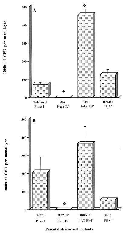
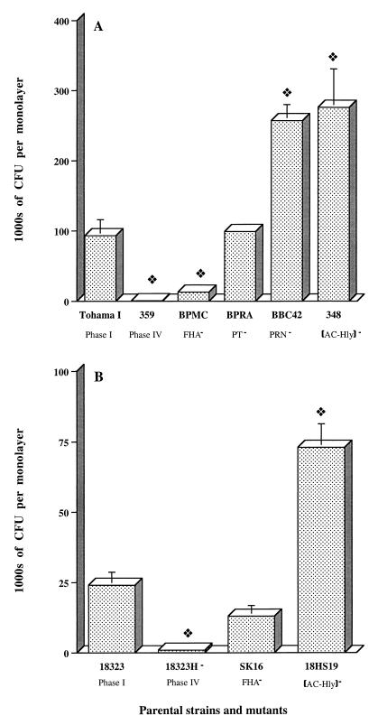
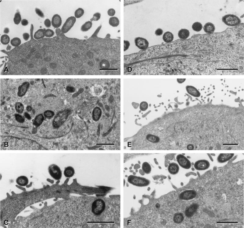
Bordetella pertussis, the agent of whooping cough, can invade and survive in several types of eukaryotic cell, including CHO, HeLa 229, and HEp-2 cells and macrophages. In this study, we analyzed bacterial invasiveness in nonrespiratory human HeLa epithelial cells and human HTE and HAE0 tracheal epithelial cells. Invasion assays and transmission electron microscopy analysis showed that B. pertussis strains invaded and survived, without multiplying, in HTE or HAE0 cells. This phenomenon was bvg regulated, but invasive properties differed between B. pertussis strains and isolates and the B. pertussis reference strain. Studies with B. pertussis mutant strains demonstrated that filamentous hemagglutinin, the major adhesin, was involved in the invasion of human tracheal epithelial cells by bacteria but not in that of HeLa cells. Fimbriae and pertussis toxin were not found to be involved. However, we found that the production of adenylate cyclase-hemolysin prevents the invasion of HeLa and HTE cells by B. pertussis because an adenylate cyclase-hemolysin-deficient mutant was found to be more invasive than the parental strain. The effect of adenylate cyclase-hemolysin was mediated by an increase in the cyclic AMP concentration in the cells. Pertactin (PRN), an adhesin, significantly inhibited the invasion of HTE cells by bacteria, probably via its interaction with adenylate cyclase-hemolysin. Isolates producing different PRNs were taken up similarly, indicating that the differences in the sequences of the PRNs produced by these isolates do not affect invasion. We concluded that filamentous hemagglutinin production favored invasion of human tracheal cells but that adenylate cyclase-hemolysin and PRN production significantly inhibited this process.


Figures

FIG. 1
FIG. 1
Invasion of HeLa 229 cell monolayers by B. pertussis parental strains and mutants. Each B. pertussis strain (7 × 10⁶ CFU) was added to a separate well of a 24-well tissue culture plate, each of which contained 7 × 10⁴ epithelial cells. The invasion of HeLa 229 cells by B. pertussis was assayed as described in Materials and Methods. The values shown are means ± SEM in thousands of CFU recovered from gentamicin-treated monolayers in three to six experiments. The diamond symbol indicates P < 0.05 versus the parental B. pertussis strain, Tohama I (A) or 18323 (B).
FIG. 2
FIG. 2
Invasion of HTE cell monolayers by B. pertussis mutants. Each B. pertussis strain (7 × 10⁶ CFU) was added to an individual well of a 24-well tissue culture plate, each of which contained 7 × 10⁴ epithelial cells. The invasion of HTE cells by B. pertussis was assayed as described in Materials and Methods. The values shown are means ± SEM in thousands of CFU recovered from gentamicin-treated monolayers in three to six experiments. The diamond symbol indicates P < 0.05 versus the parental B. pertussis strain, Tohama I (A) or 18323 (B).
FIG. 3
FIG. 3
Transmission electron micrographs showing B. pertussis within HTE cells after 1 or 3 h of coincubation. Panels: A, adherent parental strain B. pertussis Tohama I; B and C, intracellular B. pertussis Tohama I; D, adherent AC-Hly-deficient mutant B. pertussis 348; E and F, intracellular AC-Hly-deficient mutant B. pertussis 348. The lack of AC-Hly expression did not appear to render these bacteria more susceptible to damage by lysosomal enzymes. In panels C and F, note the close proximity of the bacterial outer membrane to the membrane of the endocytic vacuole. For panels A and D, the coincubation period was 1 h. For panels B, C, E, and F, the coincubation period was 3 h.
FIG. 4
FIG. 4
Invasion of HTE cell monolayers by B. pertussis isolates. Each B. pertussis strain or isolate (7 × 10⁶ CFU) was added to a separate well of a 24-well tissue culture plate each of which contained 7 × 10⁴ epithelial cells. The invasion of HTE cells by B. pertussis was assayed as described in Materials and Methods. The values shown are means ± SEM in thousands of CFU recovered from gentamicin-treated monolayers in three experiments. The diamond symbol indicates P < 0.05 compared to B. pertussis strain 18323.
FIG. 5
FIG. 5
Effect of cAMP on the invasion of HTE cells by B. pertussis. Each B. pertussis strain (7 × 10⁶ CFU) was added to a separate well of a 24-well tissue culture plate each of which contained 7 × 10⁴ epithelial cells. The invasion of HTE cells by B. pertussis was assayed as described in Materials and Methods. Forskolin was incubated with monolayers for 1 h before the addition of the AC-Hly-deficient mutant (B. pertussis 18HS19) to stimulate cAMP production. The effects of forskolin are reversible, and the concentration used (100 mM) was maintained throughout the experiment. (A) As a control, epithelial cells were incubated with (cells & forskolin) or without (control) forskolin. Results are expressed in picomoles of cAMP per 105 cells and are the means ± SEM of three independent determinations. The diamond symbol indicates P < 0.05 versus the control (cells). (B) Values are means ± SEM in thousands of CFU recovered from gentamicin-treated monolayers in three experiments. The diamond symbol indicates P < 0.05 versus B. pertussis 18323.
FIG. 6
FIG. 6
Intracellular survival of B. pertussis parental and mutant strains in human respiratory epithelial HTE cells. Each B. pertussis strain (7 × 10⁶ CFU) was added to a separate well of a 24-well tissue culture plate, each of which contained 7 × 10⁴ epithelial cells. The invasion of HTE cells by B. pertussis was assayed as described in Materials and Methods. A value of 100% corresponds to the absolute number of viable intracellular bacteria of each strain in a coincubation period of 5 h. For the 24- and 48-h points, the percentages of surviving intracellular bacteria of each strain compared to the 5-h point, independently of the parental strain, are shown. The persistence experiment was performed twice with the parental strain B. pertussis Tohama I and HTE cells and only once with the parental strain B. pertussis 18323 and the mutants. The values above the histograms for the 5-h time point correspond to the means ± SEM of the absolute number of intracellular bacteria of each strain per cell in all of the four to six experiments performed for this time point.

References

    1. Antoine R, Locht C. Roles of the disulfide bond and the carboxy-terminal region of the S1 subunit in the assembly and biosynthesis of pertussis toxin. Infect Immun. 1990;58:1518–1526. - PMC - PubMed
    1. Arico B, Gross R, Smida J, Rappuoli R. Evolutionary relationships in the genus Bordetella. Mol Microbiol. 1987;1:301–308. - PubMed
    1. Arico B, Nuti S, Scarlato V, Rappuoli R. Adhesion of Bordetella pertussis to eukaryotic cells requires a time-dependent export and maturation of filamentous hemagglutinin. Proc Natl Acad Sci USA. 1993;90:9204–9208. - PMC - PubMed
    1. Arico B, Scarlato V, Monack D M, Falkow S, Rappuoli R. Structural and genetic analysis of the bvg locus in Bordetella species. Mol Microbiol. 1991;5:2481–2491. - PubMed
    1. Boursaux-Eude C, Thiberge S, Carletti G, Guiso N. Intranasal murine model of Bordetella pertussis infection. II. Sequence variation and protection induced by a tricomponent acellular vaccine. Vaccine. 1999;17:2651–2660. - PubMed

Publication types

MeSH terms

LinkOut - more resources