. 2000 Apr;68(4):2053-60.
doi: 10.1128/IAI.68.4.2053-2060.2000.

Nonopsonic binding of type III Group B Streptococci to human neutrophils induces interleukin-8 release mediated by the p38 mitogen-activated protein kinase pathway

Affiliations

Nonopsonic binding of type III Group B Streptococci to human neutrophils induces interleukin-8 release mediated by the p38 mitogen-activated protein kinase pathway

E A Albanyan et al. Infect Immun. 2000 Apr.

Abstract

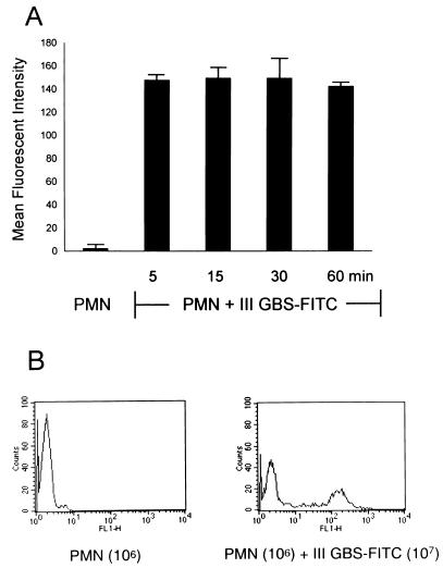
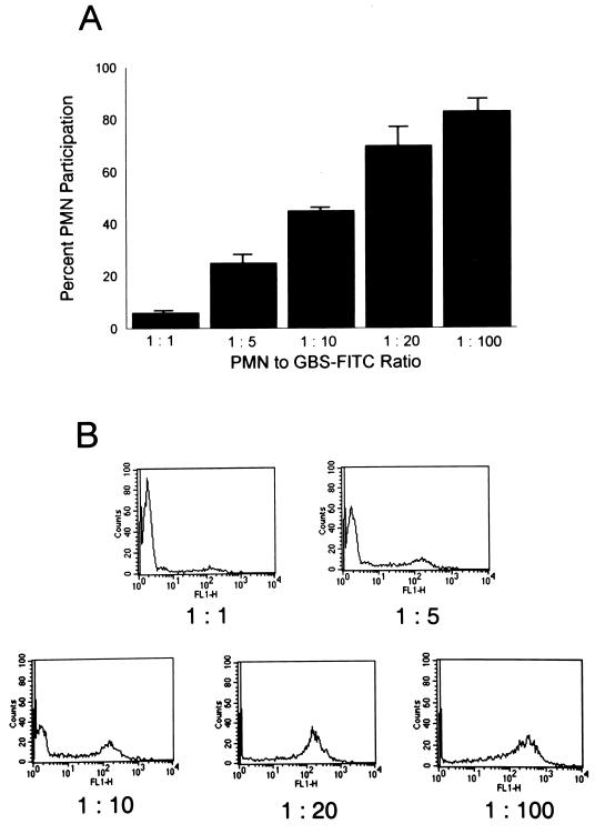
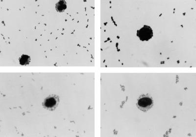
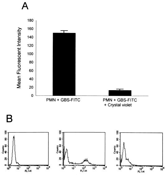
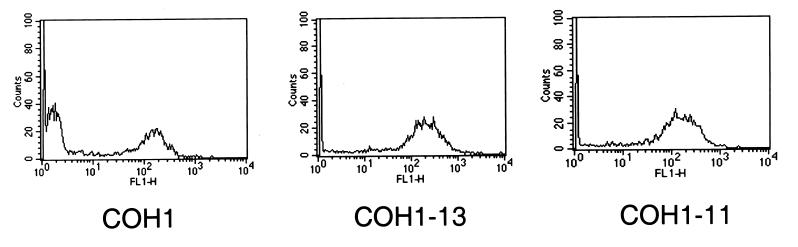
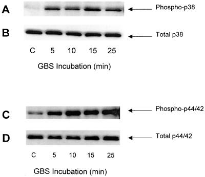
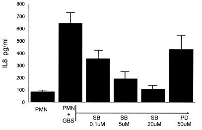
Nonopsonic interaction of host immune cells with pathogens is an important first line of defense. We hypothesized that nonopsonic recognition between type III group B streptococcus and human neutrophils would occur and that the interaction would be sufficient to trigger neutrophil activation. By using a serum-free system, it was found that heat-killed type III group B streptococci bound to neutrophils in a rapid, stable, and inoculum-dependent manner that did not result in ingestion. Transposon-derived type III strain COH1-13, which lacks capsular polysaccharide, and strain COH1-11 with capsular polysaccharide lacking terminal sialic acid demonstrated increased neutrophil binding, suggesting that capsular polysaccharide masks an underlying binding site. Experiments using monoclonal antibodies to complement receptor 1 and to the I domain or lectin site of complement receptor 3 did not inhibit binding, indicating that the complement receptors used for ingestion of opsonized group B streptococci were not required for nonopsonic binding. Nonopsonic binding resulted in rapid activation of cellular p38 and p44/42 mitogen-activated protein kinases. This interaction was not an effective trigger for superoxide production but did promote release of the proinflammatory cytokine interleukin-8. The release of interleukin-8 was markedly suppressed by the p38 mitogen-activated protein kinase inhibitor SB203580 but was only minimally suppressed by the mitogen-activated protein/extracellular signal-regulated kinase inhibitor PD98059. Thus, nonopsonic binding of type III group B streptococci to neutrophils is sufficient to initiate intracellular signaling pathways and could serve as an arm of innate immunity of particular importance to the immature host.


Figures

FIG. 1
FIG. 1
Type III GBS binds nonopsonically to PMN in a rapid and stable manner. (A) PMN (100 μl; 106) were incubated with heat-killed type III GBS-FITC (100 μl; 107 CFU) in a serum-free environment. PMN were gated according to their characteristic light scatter pattern, and data from 5,000 PMN were analyzed for increase in their MFI as an indicator of association with type III GBS-FITC. The graph shows the change in MFI of the 40% ± 3% participating PMN at this PMN-to-type III GBS ratio. This interaction occurred within 5 min, and the percentage of participating PMN and their MFI did not change, as shown by FACS analysis at 15-min intervals, for up to 1 h. (B) Histogram representative of one assay showing the MFI shift of participating PMN when incubated with type III GBS-FITC.
FIG. 2
FIG. 2
The nonopsonic interaction between PMN and type III GBS is inoculum dependent. (A) PMN (106) were incubated with increasing concentrations of heat-killed type III GBS-FITC. PMN were analyzed by flow cytometry for FITC association. The percentages of participating PMN are shown. (B) Representative family of histograms, where increasing PMN participation is evidenced by the shift in their MFI as the inoculum of type III GBS is increased.
FIG. 3
FIG. 3
Direct microscopy of the nonopsonic binding of type III GBS to PMN. PMN (106) were incubated for 5 min with heat-killed type III GBS (107 CFU). Smears of the reaction mixture were stained with NEAT STAIN (Midland Biomedical, Paulsboro, N.J.). Magnification, ×100.
FIG. 4
FIG. 4
Type III GBS is bound to but not ingested by PMN. (A) PMN (106) were incubated with heat-killed type III GBS-FITC (107 CFU), and MFI of participating PMN is shown. Crystal violet at a final concentration of 0.25 g/liter was added to quench extracellular fluorescence, and the reaction mixture was reanalyzed by flow cytometry to determine the effect on the MFI of participating PMN. The graph represents the mean ± SEM of three experiments. (B) Representative histograms showing the shift of the PMN MFI after the addition of crystal violet. The left histogram depicts the MFI of the gated PMN alone. The middle histogram shows the increase in MFI of participating PMN after incubation with type III GBS-FITC. The shift in the MFI of the PMN back to approximately baseline after the addition of crystal violet is shown in the right histogram.
FIG. 5
FIG. 5
Role of type III GBS CPS in nonopsonic binding. PMN (106) were incubated with heat-killed type III GBS-FITC using the encapsulated strain COH1 (107 CFU) or with strain COH1-13 (107 CFU), which lacks CPS, and strain COH1-11 (107 CFU), which lacks the terminal sialic acid of CPS, and were analyzed by FACS. The histograms are representative of the 44% ± 1% of PMN binding to COH1 at a PMN-to-GBS ratio of 1:10. Both COH1-13 and COH1-11 showed increased binding to PMN, with 71% ± 3% and 70% ± 4%, respectively, of gated PMN participating in the interaction at the same ratio (mean ± SEM of three experiments; P ≤ 0.02 for either mutant strain versus COH1).
FIG. 6
FIG. 6
Activation of p38 and p44/42 MAP kinases in human PMN. PMN (107/ml) were incubated with heat-killed type III GBS (108 CFU/ml) for the time intervals indicated at 37°C. Cellular proteins were separated by SDS–10 or 12% PAGE. Western blotting was performed using antibodies specific for phosphorylated p38 (A) and p44/42 (C) MAP kinases and total p38 (B) and p44/42 (D), as described in Materials and Methods. These results are representative of three similar experiments.
FIG. 7
FIG. 7
Effect of MAP kinase inhibitors on PMN IL-8 production. PMN (107/ml) were treated with the concentrations specified of SB203580 (SB) or PD98059 (PD) for 30 min. They were then incubated with heat-killed type III GBS (108 CFU/ml) for 24 h. The supernatant was separated from the cells, and the amount of IL-8 produced was determined by ELISA.

References

    1. Anderson C L, Shen L, Eicher D M, Wewers M D, Gill J K. Phagocytosis mediated by three distinct Fcγ receptor classes on human leukocytes. J Exp Med. 1990;171:1333–1345. - PMC - PubMed
    1. Antal J M, Cunningham J V, Goodrum K J. Opsonin-independent phagocytosis of group B streptococci: role of complement receptor type three. Infect Immun. 1992;60:1114–1121. - PMC - PubMed
    1. Baker, C. J., and M. S. Edwards. Group B streptococcal infections. In J. S. Remington and J. O. Klein (ed.), Infectious diseases of the fetus and newborn infant, 5th ed., in press. The W. B. Saunders Co., Philadelphia, Pa.
    1. Cantinieaux B, Hariga C, Courtoy J, Hupin J, Fondu P. Staphylococcus aureus phagocytosis. A new cytofluorometric method using FITC and paraformaldehyde. J Immunol Methods. 1989;121:203–208. - PubMed
    1. Cohen M S. Molecular events in the activation of human neutrophils for microbial killing. Clin Infect Dis. 1994;18(Suppl. 2):S170–S179. - PubMed

Publication types

MeSH terms

Substances

LinkOut - more resources