. 2000 Apr;68(4):2171-82.
doi: 10.1128/IAI.68.4.2171-2182.2000.

Role of tir and intimin in the virulence of rabbit enteropathogenic Escherichia coli serotype O103:H2

Affiliations

Role of tir and intimin in the virulence of rabbit enteropathogenic Escherichia coli serotype O103:H2

O Marchès et al. Infect Immun. 2000 Apr.

Abstract

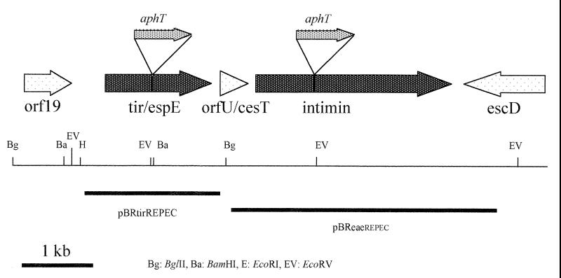
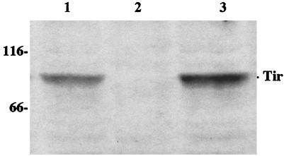
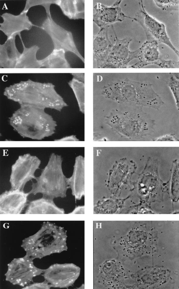
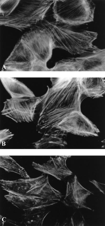
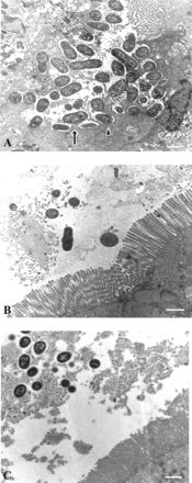
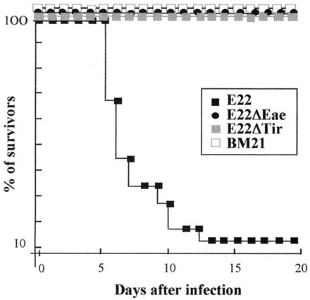
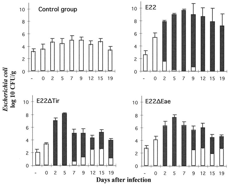
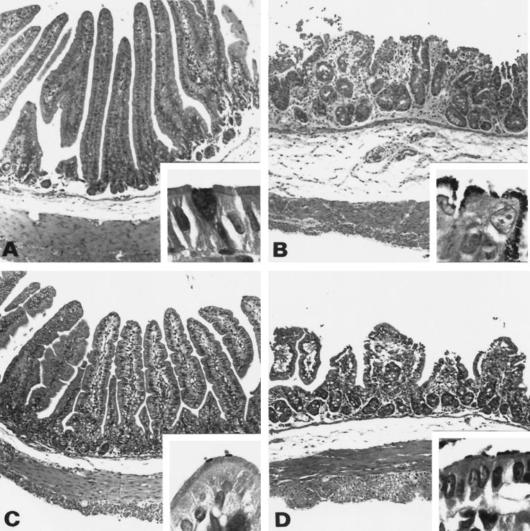
Attaching and effacing (A/E) rabbit enteropathogenic Escherichia coli (REPEC) strains belonging to serogroup O103 are an important cause of diarrhea in weaned rabbits. Like human EPEC strains, they possess the locus of enterocyte effacement clustering the genes involved in the formation of the A/E lesions. In addition, pathogenic REPEC O103 strains produce an Esp-dependent but Eae (intimin)-independent alteration of the host cell cytoskeleton characterized by the formation of focal adhesion complexes and the reorganization of the actin cytoskeleton into bundles of stress fibers. To investigate the role of intimin and its translocated coreceptor (Tir) in the pathogenicity of REPEC, we have used a newly constructed isogenic tir null mutant together with a previously described eae null mutant. When human HeLa epithelial cells were infected, the tir mutant was still able to induce the formation of stress fibers as previously reported for the eae null mutant. When the rabbit epithelial cell line RK13 was used, REPEC O103 produced a classical fluorescent actin staining (FAS) effect, whereas both the eae and tir mutants were FAS negative. In a rabbit ligated ileal loop model, neither mutant was able to induce A/E lesions. In contrast to the parental strain, which intimately adhered to the enterocytes and destroyed the brush border microvilli, bacteria of both mutants were clustered in the mucus without reaching and damaging the microvilli. The role of intimin and Tir was then analyzed in vivo by oral inoculation of weaned rabbits. Although both mutants were still present in the intestinal flora of the rabbits 3 weeks after oral inoculation, neither mutant strain induced any clinical signs or significant weight loss in the inoculated rabbits whereas the parental strain caused the death of 90% of the inoculated rabbits. Nevertheless, an inflammatory infiltrate was present in the lamina propria of the rabbits infected with both mutants, with an inflammatory response greater for the eae null mutant. In conclusion, we have confirmed the role of intimin in virulence, and we have shown, for the first time, that Tir is also a key factor in vivo for pathogenicity.


Figures

FIG. 1
FIG. 1
Genetic and restriction maps of the REPEC O103 LEE locus, encoding Tir and intimin. Sites of insertional mutation in tir and eae and fragments cloned in plasmid pBRSK giving pBReaeREPEC and pBRtirREPEC and used to complement mutant strains E22ΔEae and E22ΔTir are shown.
FIG. 2
FIG. 2
Western blot of total cell lysates of E22ΔEae(pBReaeREPEC) (lane 1), E22ΔEae (lane 2), and E22 (lane 3) grown in Dulbecco's modified Eagle's medium. Samples were resolved by SDS–8% PAGE and transferred to PVDF membranes. The membranes were probed with a serum raised against an Eae–maltose-binding protein fusion protein. Molecular mass markers are given in kilodaltons on the left.
FIG. 3
FIG. 3
Western blot of the Triton X-100-insoluble fraction of HeLa cells infected for 4 h with E22 (lane 1), E22ΔTir (lane 2), and E22ΔTir(pBRtirREPEC) (lane 3). Samples were resolved by SDS–8% PAGE and transferred to PVDF membranes. The membranes were probed with an antiphosphotyrosine monoclonal antibody. Molecular mass markers are given in kilodaltons on the left.
FIG. 4
FIG. 4
REPEC O103 strains E22ΔEae (A and B) and E22ΔTir (E and F) lost their ability to focus F-actin beneath their adhesion sites. The FAS response was restored upon trans complementation of each mutant with its respective parental gene eae (C and D) or tir (G and H). RK13 cells were infected for 4 h with the challenged strains, and then the monolayers were washed, fixed, and permeabilized and F-actin was labeled with rhodamine-phalloidin. Fluorescence (A, C, E, and G) and corresponding phase-contrast (B, D, F, and H) micrographs were taken with a Leica X500 microscope.
FIG. 5
FIG. 5
Alteration of actin cytoskeleton in HeLa cells 36 h after interaction with E22 (wild type) (A), E22ΔTir (B), and a K-12 laboratory strain (C). Both strains E22 (A) and E22ΔTir (B) induced a cell size increase with a multiplication of actin stress fibers characteristic of the CPE. Actin was labeled with rhodamine-phalloidine, and samples were observed with a Leica X300 microscope.
FIG. 6
FIG. 6
Transmission electron microscopy of rabbit ligated intestinal loops inoculated with E22 (A), E22ΔEae (B), and E22ΔTir (C). E22 (A) adhered in a diffuse pattern to the enterocytes and induced typical A/E lesions characterized by an intimate attachment of bacteria to the enterocyte surface on pedestal structures (arrow) or cup-like structures (arrowhead) and associated with a destruction of surrounding microvilli. In contrast, no A/E lesions were observed with E22ΔEae (B) and E22ΔTir (C). Brush border microvilli were intact, and adherent bacteria stayed clustered in the mucus without reaching the enterocytes (B and C). Samples were analyzed 24 h after infection. Bars, 1 μm.
FIG. 7
FIG. 7
Percentages of rabbits that survived infection after oral inoculation of E22, E22ΔEae, E22ΔTir, and BM21.
FIG. 8
FIG. 8
Kinetics of fecal E. coli shedding in rabbits inoculated orally with wild-type rabbit REPEC strain E22, mutant strains E22ΔEae and E22ΔTir, and laboratory strain K-12 BM21. Solid bars represent the relative proportions of inoculated strains (E22, E22ΔEae, and E22ΔTir) compared to the normal E. coli population (white bars). The laboratory strain BM21 was not recovered from the feces of the control group throughout the experiment.
FIG. 9
FIG. 9
Rabbit distal ileum tissue sections 5 days after inoculation. (A) Avirulent strain BM21; (B) E22; (C) E22ΔTir; (D) E22ΔEae. At low-power magnification (×100; hematoxylin-eosin staining), micrographs show the variation of the atrophy level of small intestinal villi, epithelial exfoliation, inflammatory infiltrate of the lamina propria, and edematous aspects of the submucosa, depending on the strain inoculated. (A) Normal histological structure of the small bowel villi. (B) Severely atrophic and fused villi, epithelial exfoliation, and marked inflammatory infiltrate of the lamina propria and submucosa. (C) Slight atrophy of the villi and inflammatory infiltrate of the proprial core of the villi. (D) Pronounced atrophy associated with polymorphous infiltrate extending to the oedematous submucosa. Higher magnification (insets; ×1,000; Gram staining) shows the normal appearance of the columnar enterocyte with a prominent brush border (A); for both mutants, the gram-negative bacilli observed are just at the extremity of the microvilli on the surface of the brush border, which seems to be relatively well preserved and not effaced (C and D). The enterocytes in the panel B inset are irregular and rounded up, and the microvillous border is indistinct and covered by a heavy layer of prominent gram-negative bacilli.

References

    1. Abe A, Heczko U, Hegele R G, Finlay B B. Two enteropathogenic Escherichia coli type III secreted proteins, EspA and EspB, are virulence factors. J Exp Med. 1998;188:1907–1916. - PMC - PubMed
    1. Adams L M, Simmons C P, Rezmann L, Strugnell R A, Robins-Browne R M. Identification and characterization of a K88- and CS31A-like operon of a rabbit enteropathogenic Escherichia coli strain which encodes fimbriae involved in the colonization of rabbit intestine. Infect Immun. 1997;65:5222–5230. - PMC - PubMed
    1. An H, Fairbrother J M, Dubreuil J D, Harel J. Cloning and characterization of the eae gene from a dog attaching and effacing Escherichia coli strain 4221. FEMS Microbiol Lett. 1997;148:239–245. - PubMed
    1. Bieber D, Ramer S W, Wu C Y, Murray W J, Tobe T, Fernandez R, Schoolnik G K. Type IV pili, transient bacterial aggregates, and virulence of enteropathogenic Escherichia coli. Science. 1998;280:2114–2118. - PubMed
    1. Blanco J E, Blanco M, Blanco J, Mora A, Balaguer L, Mourino M, Juarez A, Jansen W H. O serogroups, biotypes, and eae genes in Escherichia coli strains isolated from diarrheic and healthy rabbits. J Clin Microbiol. 1996;34:3101–3107. - PMC - PubMed

Publication types

MeSH terms

Associated data