. 2000 Apr;68(4):2276-85.
doi: 10.1128/IAI.68.4.2276-2285.2000.

The leptospiral major outer membrane protein LipL32 is a lipoprotein expressed during mammalian infection

Affiliations

The leptospiral major outer membrane protein LipL32 is a lipoprotein expressed during mammalian infection

D A Haake et al. Infect Immun. 2000 Apr.

Abstract

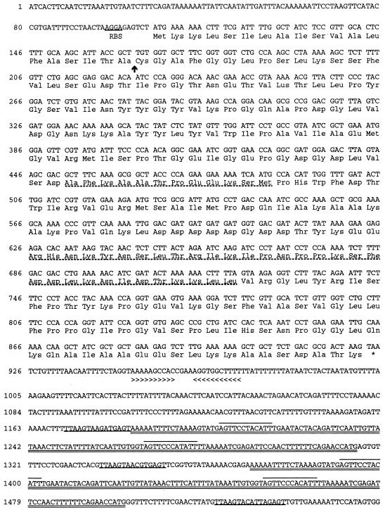
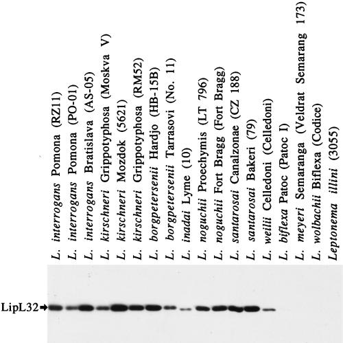
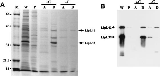
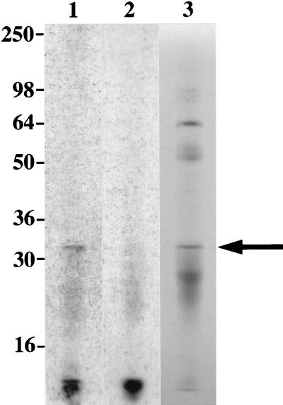
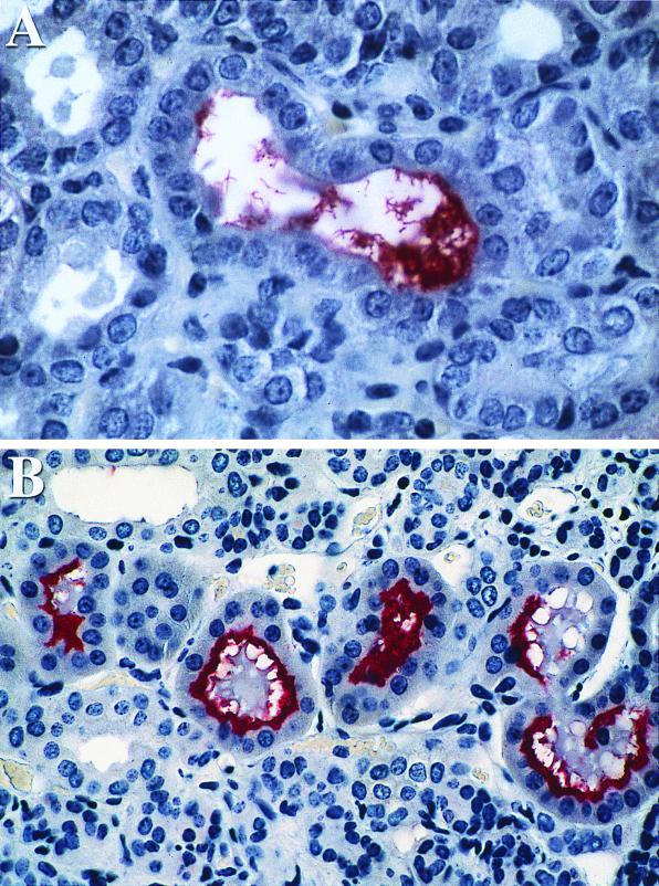
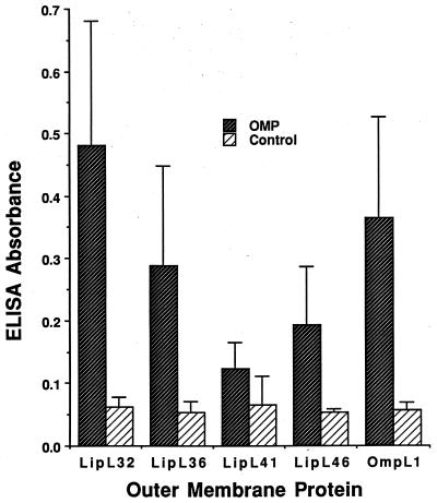
We report the cloning of the gene encoding the 32-kDa lipoprotein, designated LipL32, the most prominent protein in the leptospiral protein profile. We obtained the N-terminal amino acid sequence of a staphylococcal V8 proteolytic-digest fragment to design an oligonucleotide probe. A Lambda-Zap II library containing EcoRI fragments of Leptospira kirschneri DNA was screened, and a 5.0-kb DNA fragment which contained the entire structural lipL32 gene was identified. Several lines of evidence indicate that LipL32 is lipid modified in a manner similar to that of other procaryotic lipoproteins. The deduced amino acid sequence of LipL32 would encode a 272-amino-acid polypeptide with a 19-amino-acid signal peptide, followed by a lipoprotein signal peptidase cleavage site. LipL32 is intrinsically labeled during incubation of L. kirschneri in media containing [(3)H]palmitate. The linkage of palmitate and the amino-terminal cysteine of LipL32 is acid labile. LipL32 is completely solubilized by Triton X-114 extraction of L. kirschneri; phase separation results in partitioning of LipL32 exclusively into the hydrophobic, detergent phase, indicating that it is a component of the leptospiral outer membrane. CaCl(2) (20 mM) must be present during phase separation for recovery of LipL32. LipL32 is expressed not only during cultivation but also during mammalian infection. Immunohistochemistry demonstrated intense LipL32 reactivity with L. kirschneri infecting proximal tubules of hamster kidneys. LipL32 is also a prominent immunogen during human leptospirosis. The sequence and expression of LipL32 is highly conserved among pathogenic Leptospira species. These findings indicate that LipL32 may be important in the pathogenesis, diagnosis, and prevention of leptospirosis.


Figures

FIG. 1
FIG. 1
Nucleotide sequence and deduced amino acid sequence of lipL32. The putative ribosome-binding site (RBS) is shown. The putative signal peptidase cleavage site is indicated by an arrow. The amino acid sequences obtained from the staphylococcal V8 protease digestion of the native protein are shown (broken underline). The location of the TAA stop codon is indicated by an asterisk. An inverted repeat is indicated by arrowheads. Repeated sequences following the lipL32 gene include a shorter sequence repeated three times (underlined), flanking a longer sequence repeated twice (double underlined). The longer sequences contain another short sequence which is repeated four times (overlined).
FIG. 2
FIG. 2
Phylogenetic tree based on lipL32 nucleotide sequence differences. L. kirschneri serovar grippotyphosa, L. interrogans serovar pomona, and L. interrogans serovar lai formed one monophyletic group; a second group was formed by L. borgpetersenii serovar hardjo and L. santarosai serovar tropica, while L. noguchii serovar fortbragg segregated separately. Sequences were analyzed by using the PAUP software package (version 3.1; D. Swafford, Smithsonian Institution, Washington, D.C.). Horizontal lengths are proportional to nucleotide step differences (indicated above the lines).
FIG. 3
FIG. 3
Immunoblot of a panel of Leptospira species obtained by using LipL32 antiserum. LipL32 antiserum detected a single band in each of the lanes containing pathogenic Leptospira species. LipL32 expression is highly conserved among the pathogenic Leptospira species, L. interrogans, L. kirschneri, L. borgpetersenii, L. inadai, L. noguchii, L. santarosai, and L. weilii. L. biflexa, L. meyeri, and L. wolbachii are nonpathogenic species, as is the related organism Leptonema illini.
FIG. 4
FIG. 4
Behavior of LipL32 and other leptospiral proteins in Triton X-114. Triton X-114 fractions of L. kirschneri organisms were separated by SDS-PAGE and stained with Coomassie brilliant blue (A) or probed with either a combination of LipL32 and LipL41 antisera (B). Fractions analyzed were the whole organism (W), Triton X-114-insoluble pellet (P), and aqueous-phase (A) or detergent-phase (D) material with (+C) or without (−C) 20 mM CaCl2. LipL32, but not LipL41, requires CaCl2 to avoid proteolytic degradation during Triton X-114 phase partitioning.
FIG. 5
FIG. 5
LipL32 is acylated by L. kirschneri. LipL32 was isolated from L. kirschneri organisms intrinsically labeled with [3H]palmitate by extraction with Triton X-100 and immunoprecipitation with LipL32 antiserum. Lanes 1, autoradiogram of immunoprecipitated LipL32 without treatment of SDS-PAGE gel with acetic acid; 2, autoradiogram of immunoprecipitated LipL32 after treatment of SDS-PAGE gel with acetic acid; 3, immunoprecipitated LipL32 stained with Coomassie brilliant blue. Acetic acid treatment of SDS-PAGE gel results in a loss of 3H label, a finding consistent with an acid-labile bond between the labeled palmitate and the N-terminal cysteine of LipL32. Locations of molecular size standards are shown (in kilodaltons) on the left.
FIG. 6
FIG. 6
Immunohistochemistry of kidney tissue obtained at 10 days (A) and 28 days (B) postinfection with virulent L. kirschneri by using LipL32 antiserum. Antigen was detected on leptospires within the renal tubular lumen at both time points. The increased reactivity at 28 days after challenge is consistent with a higher burden of organisms.
FIG. 7
FIG. 7
Antibodies to L. kirschneri outer membrane proteins (OMPs) in humans with leptospirosis. Mean ELISA readings to leptospiral outer membrane proteins in the convalescent sera from five leptospirosis patients from Barbados are shown. Control readings are from wells with sera but without antigen. Standard deviations are shown by the error bars. Differences between wells with or without antigen were significant for all five membrane proteins (P < 0.01), but there was a greater degree of statistical significance for OmpL1 (P < 0.001) and LipL32 (P < 0.0001).

References

    1. Altschul S F, Madden T L, Schaffer A A, Zhang J, Zhang Z, Miller W, Lipman D J. Gapped BLAST and PSI-BLAST: a new generation of protein database search programs. Nucleic Acids Res. 1997;25:3389–3402. - PMC - PubMed
    1. Auran N E, Johnson R C, Ritzi D M. Isolation of the outer sheath of Leptospira and its immunogenic properties in hamsters. Infect Immun. 1972;5:968–975. - PMC - PubMed
    1. Barnett J K, Barnett D, Bolin C A, Summers T A, Wagar E A, Cheville N F, Hartskeerl R A, Haake D A. Expression and distribution of leptospiral outer membrane components during renal infection of hamsters. Infect Immun. 1999;67:853–861. - PMC - PubMed
    1. Bergstrom S, Bundoc V G, Barbour A G. Molecular analysis of linear plasmid-encoded major surface proteins, OspA and OspB, of the Lyme disease spirochaete Borrelia burgdorferi. Mol Microbiol. 1989;3:479–486. - PubMed
    1. Bolin C A, Cassells J A, Zuerner R L, Trueba G. Effect of vaccination with a monovalent Leptospira interrogans serovar hardjo type hardjo-bovis vaccine on type hardjo-bovis infection of cattle. Am J Vet Res. 1991;52:1639–1643. - PubMed

Publication types

MeSH terms

LinkOut - more resources