

doi: 10.1128/IAI.68.4.2379-2385.2000.
Ultrastructural analysis of developmental events in Chlamydia pneumoniae-infected cells
Affiliations
- PMID: 10722649
- PMCID: PMC97433
- DOI: 10.1128/IAI.68.4.2379-2385.2000
Item in Clipboard
Ultrastructural analysis of developmental events in Chlamydia pneumoniae-infected cells
Infect Immun.
2000 Apr.
Abstract
Chlamydia pneumoniae is an obligate intracellular parasite with a developmental cycle believed to be common to all members of the genus Chlamydia. We present a detailed description based on transmission and scanning electron microscopy of temporal events and inclusion structures throughout the C. pneumoniae AR-39 developmental cycle.
Figures
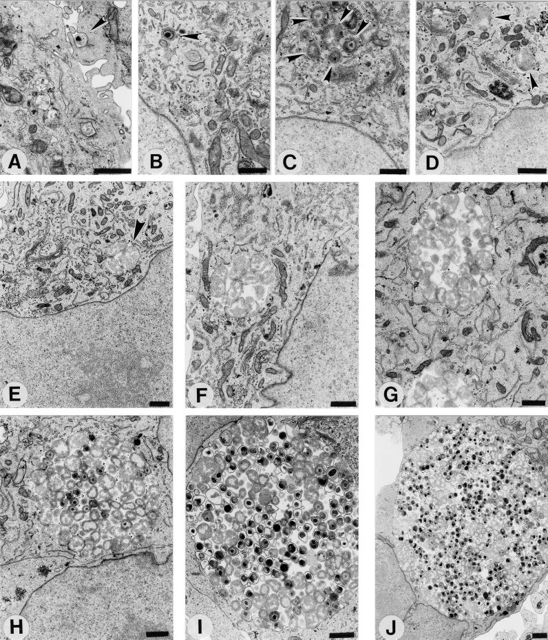
The C. pneumoniae AR-39 developmental cycle in HeLa 229 cells. Infected cells were processed for transmission electron microscopy at 0 (A), 2 (B), 8 (C), 12 (D), 19 (E), 24 (F), 36 (G), 48 (H), 60 (I), and 72 (J) h postinfection. Arrowheads indicate intracellular chlamydiae. Scale bars = 1 μm.
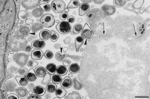
Transmission electron micrograph of a C. pneumoniae inclusion at 72 h postinfection showing details of the mature EBs, including the pear shape as well as the presence of miniature bodies (arrowheads) within the EBs' cytoplasms. Arrows indicate sites suggestive of septation and possible generation of C. pneumoniae RBs by budding from a giant RB located in the lower right corner. Scale bar = 0.5 μm.
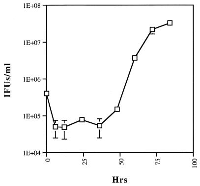
A one-step growth curve of C. pneumoniae AR-39 shows an initial 10-fold decrease in the number of infectious units and the maintenance of this level up to 36 h postinfection. The number of inclusion-forming units (IFUs) increases by 48 h postinfection, when the differentiation of RBs to EBs is initiated. Differentiation continues up to 84 h postinfection.
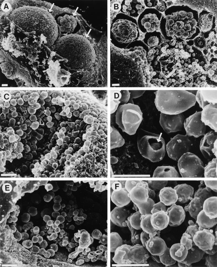
Scanning electron micrographs of C. pneumoniae AR-39 and C. trachomatis L2 inclusions in fractured host cells. (A and B) Presence of several C. pneumoniae inclusions within individual infected HeLa cells (arrows). Each of these inclusions appears to be at a different stage of maturity. Scale bars = 1 μm. The remaining panels emphasize the differences in shape between EBs of C. pneumoniae (C and D) and those of C. trachomatis (E and F) and their distribution inside the inclusion, as well as the different structures of inclusions between these two chlamydial species. Scanning electron microscopy confirmed the pear shape of C. pneumoniae EBs, and a break in an EB (arrow in panel D) reveals the presence of a large periplasmic space between the cytoplasmic and the outer membranes of the EBs. Scale bars = 0.5 μm.
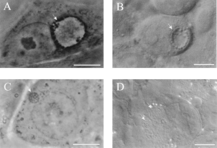
Phase- and Nomarski differential-interference-contrast images of C. trachomatis (A and B, respectively) and C. pneumoniae (C and D) inclusions. In comparison to inclusions of C. trachomatis obtained 18 h postinfection, C. pneumoniae inclusions at 36 h postinfection differ in shape and size, although both chlamydiae reach similar stages of their development at these time points postinfection, characterized by the presence of dividing RBs. This difference is most apparent in the presence of clear fluid-filled centers in C. trachomatis inclusions, since C. pneumoniae inclusions are filled with RBs. Scale bars = 10 μm.
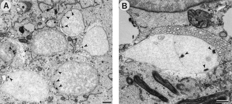
Transmission electron micrograph of C. pneumoniae-infected cells treated with ampicillin. (A) Large, abnormal RBs displaying single-cell or no cell division and multiple electron-dense sites, possibly representing nucleoid condensation, near the chlamydial cell wall (arrowheads). (B) Vesicles of unknown function and origin surrounded some giant RBs. Scale bars = 0.5 μm.
References
-
- Carter M W, Al-Mahdawi S A H, Giles I G, Treharne J D, Ward M E, Clarke I N. Nucleotide sequence and taxonomic value of the major outer membrane protein gene of Chlamydia pneumoniae IOL-207. J Gen Microbiol. 1991;137:465–475. - PubMed
MeSH terms
LinkOut - more resources
Full Text Sources
Other Literature Sources
Research Materials
