. 2000 Mar 20;148(6):1107-13.
doi: 10.1083/jcb.148.6.1107.

Reverse transcriptase activity in mature spermatozoa of mouse

Affiliations

Reverse transcriptase activity in mature spermatozoa of mouse

R Giordano et al. J Cell Biol. .

Abstract

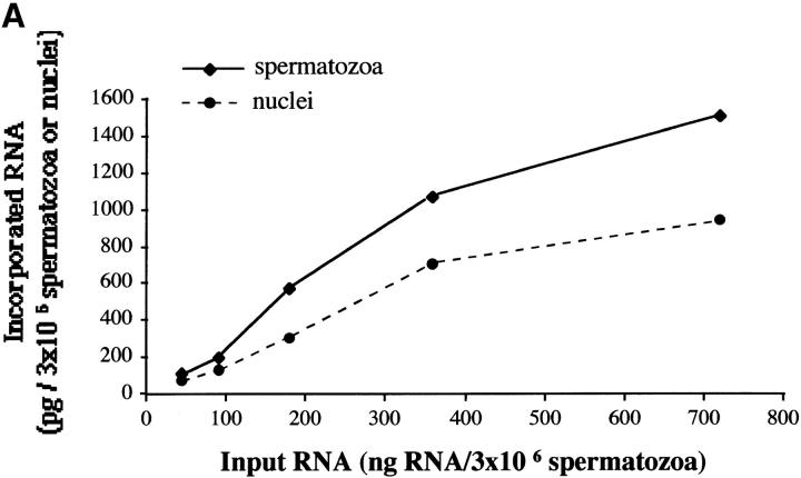
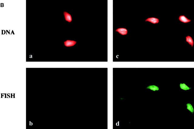
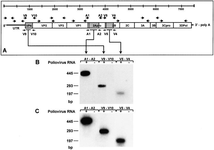
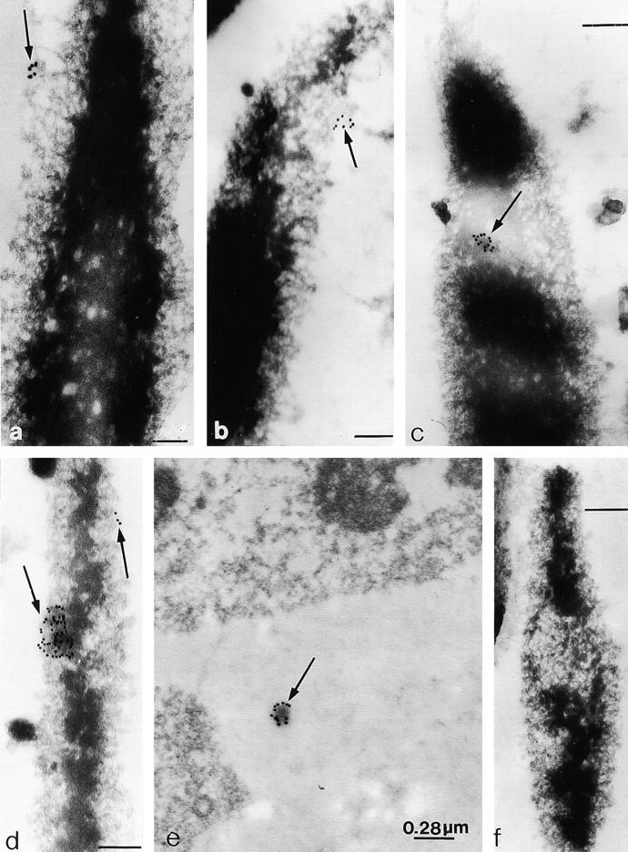
We show here that a reverse transcriptase (RT) activity is present in murine epididymal spermatozoa. Sperm cells incubated with human poliovirus RNA can take up exogenous RNA molecules and internalize them in nuclei. Direct PCR amplification of DNA extracted from RNA-incubated spermatozoa indicate that poliovirus RNA is reverse-transcribed in cDNA fragments. PCR analysis of two-cell embryos shows that poliovirus RNA-challenged spermatozoa transfer retrotranscribed cDNA molecules into eggs during in vitro fertilization. Finally, RT molecules can be visualized on sperm nuclear scaffolds by immunogold electron microscopy. These results, therefore, reveal a novel metabolic function in spermatozoa, which may play a role during early embryonic development.


Figures

Figure 1
Figure 1
(A) Association of end-labeled poliovirus RNA with mouse epididymal sperm cells. Spermatozoa aliquots were incubated with 45, 90, 180, 360, and 720 ng of poliovirus RNA. After 30 min, spermatozoa from each incubation mixture were washed and divided in two aliquots. The first one was dissolved in soluene/toluene scintillation cocktail and counted to measure the RNA uptake; the second aliquot was used for nuclei purification, then treated and counted as for whole cells to measure nuclear internalization. (B) FISH localization of poliovirus RNA in isolated nuclei from RNA-challenged spermatozoa. (a and c) Sperm nuclei stained with DAPI and pseudocolored in red; (b) FISH experiment with nuclei from spermatozoa incubated with buffer only; (d) FISH experiment with nuclei from spermatozoa incubated with poliovirus RNA. The signal associated with the biotinylated probe is pseudocolored in green.
Figure 1
Figure 1
(A) Association of end-labeled poliovirus RNA with mouse epididymal sperm cells. Spermatozoa aliquots were incubated with 45, 90, 180, 360, and 720 ng of poliovirus RNA. After 30 min, spermatozoa from each incubation mixture were washed and divided in two aliquots. The first one was dissolved in soluene/toluene scintillation cocktail and counted to measure the RNA uptake; the second aliquot was used for nuclei purification, then treated and counted as for whole cells to measure nuclear internalization. (B) FISH localization of poliovirus RNA in isolated nuclei from RNA-challenged spermatozoa. (a and c) Sperm nuclei stained with DAPI and pseudocolored in red; (b) FISH experiment with nuclei from spermatozoa incubated with buffer only; (d) FISH experiment with nuclei from spermatozoa incubated with poliovirus RNA. The signal associated with the biotinylated probe is pseudocolored in green.
Figure 2
Figure 2
PCR amplification of poliovirus cDNA copies in sperm cells and two-cell embryos after incubation with poliovirus RNA. (A) Map of the 7,433-nt poliovirus RNA chromosome. Solid boxes indicate regions of the viral genome that were found to be amplified in the DNA extracted from both poliovirus RNA-incubated spermatozoa and from two-cell embryos. Arrows indicate the oligonucleotide combinations used to amplify the viral chromosome. Viral cDNAs fragments were PCR-amplified in DNA samples extracted from: (B) poliovirus RNA-challenged (+), but not from buffer-incubated (−) spermatozoa, and (C) two-cell embryos after egg fertilization with poliovirus RNA-incubated (+), but not with buffer-incubated (−), spermatozoa. Amplified cDNAs were visualized by hybridization with specific internal oligonucleotide probes.
Figure 3
Figure 3
Localization of RT molecules (arrowed) on sperm nuclear scaffolds by immunogold electron microscopy. (a–d) Sperm nuclear scaffolds; (e) HIV-infected T-lymphocyte; (f) scaffolds incubated with secondary antibody conjugated with colloidal gold particles. Bars: (a, c, d, and f) 32 μm; (b) 25 μm.

References

    1. Balhorn R., Gladhill B.L., Wyrobek A.J. Mouse sperm chromatin proteinsquantitative isolation and partial characterization. Biochemistry. 1977;16:4074–4080. - PubMed
    1. Baron M.H., Baltimore D. Antibodies against the chemically synthesized genome-linked protein of poliovirus react with native virus-specific proteins. Cell. 1982;28:395–404. - PubMed
    1. Boyle A.L., Ballard S.G., Ward D.C. Differential distribution of long and short interspersed element sequences in the mouse genomechromosome karyotyping by fluorescence in situ hybridization. Proc. Natl. Acad. Sci. USA. 1990;87:7757–7761. - PMC - PubMed
    1. Carlemalm E., Villiger W., Hobot J.A., Acetarin J.D., Kellenberger E. Low temperature embedding with Lowicryl resinstwo new formulations and some applications. J. Microsc. 1985;140:55–63. - PubMed
    1. Eisenhauer K.M., Gerstein R.M., Chiu C.P., Conti M., Hsueh A.J. Telomerase activity in female and male rat germ cells undergoing meiosis and in early embryos. Biol. Reprod. 1997;56:1120–1125. - PubMed

Publication types