. 2000 Mar 20;148(6):1165-76.
doi: 10.1083/jcb.148.6.1165.

Ubinuclein, a novel nuclear protein interacting with cellular and viral transcription factors

Affiliations

Ubinuclein, a novel nuclear protein interacting with cellular and viral transcription factors

S Aho et al. J Cell Biol. .

Abstract

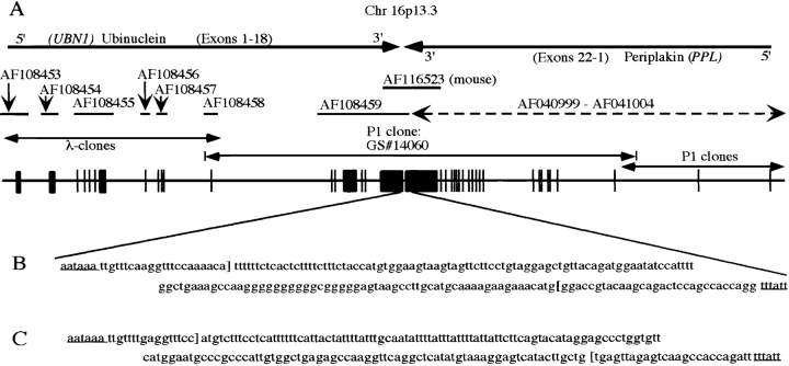
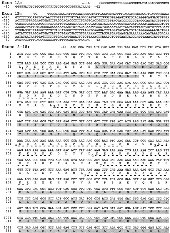
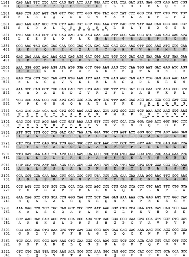
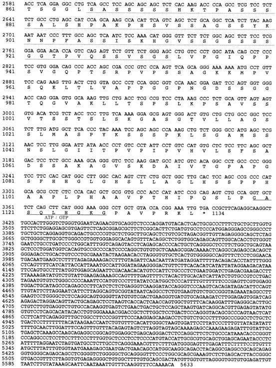
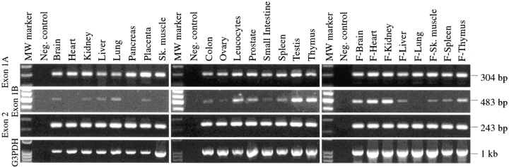
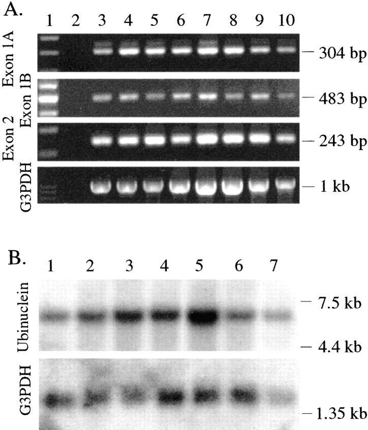
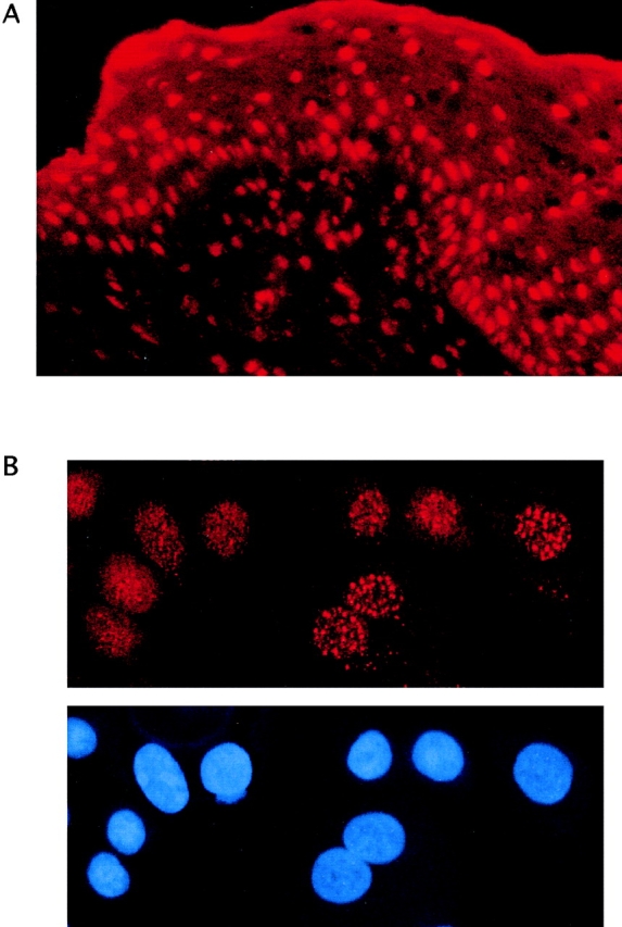
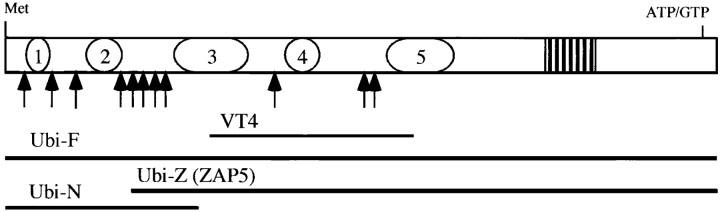
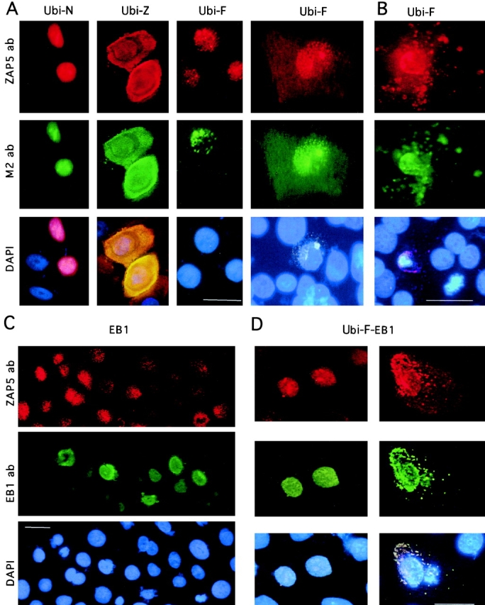
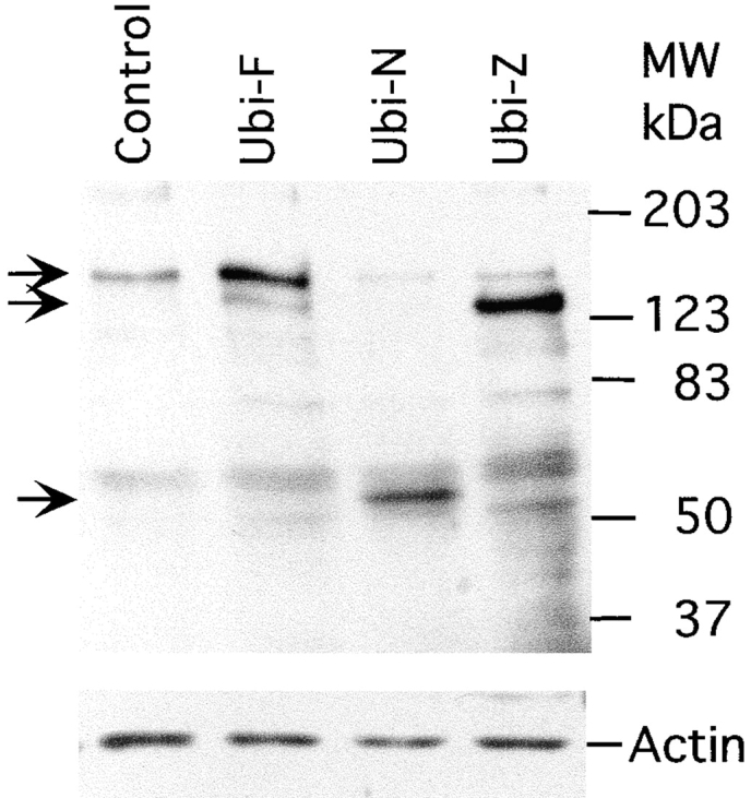
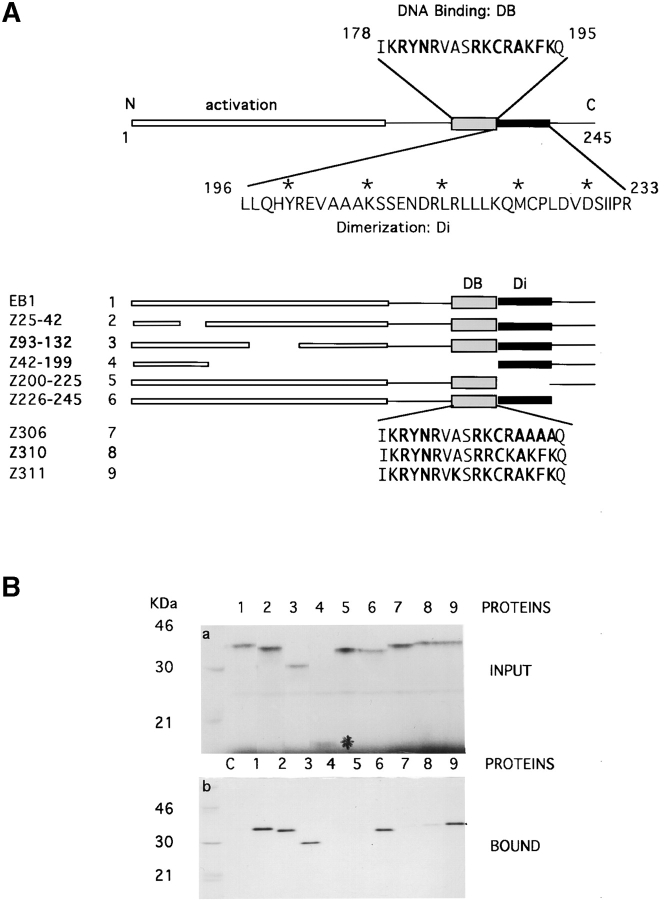
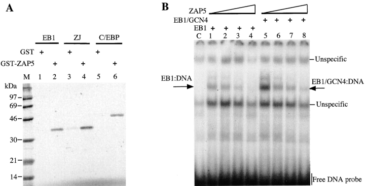
The major target tissues for Epstein-Barr virus (EBV) infection are B lymphocytes and epithelial cells of the oropharyngeal zone. The product of the EBV BZLF1 early gene, EB1, a member of the basic leucine-zipper family of transcription factors, interacts with both viral and cellular promoters and transcription factors, modulating the reactivation of latent EBV infection. Here, we characterize a novel cellular protein interacting with the basic domains of EB1 and c-Jun, and competing of their binding to the AP1 consensus site. The transcript is present in a wide variety of human adult, fetal, and tumor tissues, and the protein is detected in the nuclei throughout the human epidermis and as either grainy or punctuate nuclear staining in the cultured keratinocytes. The overexpression of tagged cDNA constructs in keratinocytes revealed that the NH(2) terminus is essential for the nuclear localization, while the central domain is responsible for the interaction with EB1 and for the phenotype of transfected keratinocytes similar to terminal differentiation. The gene was identified in tail-to-tail orientation with the periplakin gene (PPL) in human chromosome 16p13.3 and in a syntenic region in mouse chromosome 16. We designated this novel ubiquitously expressed nuclear protein as ubinuclein and the corresponding gene as UBN1.


Figures

Figure 1
Figure 1
Genomic organization of the periplakin/ubinuclein locus on human chromosome 16p13.3. A, Sequencing of the human genomic P1 clone, GS14060, containing PPL disclosed the presence of a novel gene, UBN1. The orientation of each gene is shown on top, and the accession numbers for the DNA sequences available from the GenBank/EMBL/DDBJ are denoted in the middle. The exons drawn and placed in scale are shown as black squares. B, Human UBN1 and PPL genes were found to reside in tail-to-tail orientation. The distance between the corresponding polyadenylation signals (AATAAA and TTTATT, underlined) is 194 bp. The ends of the cDNAs are indicated by brackets. C, The DNA sequencing of a mouse genomic λ-clone revealed that the arrangement of the mouse Ubn1/Ppl locus on the syntenic region of mouse chromosome 16 was similar to that of the human locus, and the distance between the polyadenylation signals is 197 bp.
Figure 2
Figure 2
Nucleotide sequence of the cDNA encoding ubinuclein and the predicted amino acid sequence. Numbering of the cDNA sequence begins from the first nucleotide of the putative translation initiation codon, ATG, which is preceded by an in-frame stop codon, TAA, only six triplets upstream (underlined). Two alternative 5′-ends probably due to the multiple promoter usage, denoted Exon 1A and Exon 1B, were discovered. Five nucleolin-type acidic regions are highlighted by shading. Repeats of basic amino acids with homology to nuclear localization signals are underlined by dashed lines and numbered (1–11). The serine-rich region is denoted with dotted underlining and the ATP/GTP consensus binding sequence (amino acids 1119–1127) is underlined. The open reading frame encoding a polypeptide of 1134 amino acid residues terminates in a stop codon (indicated by an asterisk), which is followed by a consensus polyadenylation signal (AATAAA, underlined) at the end of the 2280-bp 3′-UTR. These sequence data are available from GenBank/EMBL/DDBJ under accession numbers AF108460 and AF108461.
Figure 2
Figure 2
Nucleotide sequence of the cDNA encoding ubinuclein and the predicted amino acid sequence. Numbering of the cDNA sequence begins from the first nucleotide of the putative translation initiation codon, ATG, which is preceded by an in-frame stop codon, TAA, only six triplets upstream (underlined). Two alternative 5′-ends probably due to the multiple promoter usage, denoted Exon 1A and Exon 1B, were discovered. Five nucleolin-type acidic regions are highlighted by shading. Repeats of basic amino acids with homology to nuclear localization signals are underlined by dashed lines and numbered (1–11). The serine-rich region is denoted with dotted underlining and the ATP/GTP consensus binding sequence (amino acids 1119–1127) is underlined. The open reading frame encoding a polypeptide of 1134 amino acid residues terminates in a stop codon (indicated by an asterisk), which is followed by a consensus polyadenylation signal (AATAAA, underlined) at the end of the 2280-bp 3′-UTR. These sequence data are available from GenBank/EMBL/DDBJ under accession numbers AF108460 and AF108461.
Figure 3
Figure 3
Expression of ubinuclein transcripts in human adult and fetal tissues. The multiple tissue cDNA panels I and II (first two panels), and human fetal panel (last panel) were used as PCR templates. PCR primers specific for exon 1A–exon 2, exon 1B–exon 2, and exon 2 of ubinuclein, and for G3PDH as an internal standard, were used. cDNAs were derived from polyA+ RNA isolated from tissues indicated on the top and the quantity was normalized against the transcripts of several housekeeping genes. In most tissues, transcripts with exon 1A–exon 2-specific primers were detected at the equal level, whereas a transcript containing exon 1B was less abundant.
Figure 4
Figure 4
Expression of ubinuclein transcript in human tumor tissues. A, Tumor tissue cDNA panel was used as a PCR template as described in Fig. 3. Lane 1, Molecular weight marker, 100-bp ladder; lane 2, negative control; lane 3, colon adenocarcinoma (GI-112); lane 4, colon adenocarcinoma (CX-1); lane 5, pancreatic adenocarcinoma (GI-103); lane 6, prostatic adenocarcinoma (PC-3); lane 7, lung carcinoma (GI-117); lane 8, lung carcinoma (LX-1); lane 9, breast carcinoma (GI-101); lane 10, ovarian carcinoma (GI-102). B, Northern blot analysis of human mRNA. Hybridization of the human cancer cell line multiple tissue Northern blot with the ubinuclein-specific probe revealed a transcript of 7 kb. G3PDH-specific probe was used as a control. Lane 1, Promyelocytic leukemia HL-60; lane 2, HeLa cells S3; lane 3, chronic myelogenous leukemia k-562; lane 4, lymphoblastic leukemia MOLT-4; lane 5, colorectal adenocarcinoma SW40; lane 6, lung carcinoma A549; lane 7, melanoma G361.
Figure 5
Figure 5
Nuclear localization of the ubinuclein protein. IIF of a newborn foreskin (A) and primary foreskin keratinocytes in culture (B). Top of B shows Ubi-Z/ZAP5 antibody staining visualized with Texas red, whereas nuclei are shown by DAPI in the bottom panel.
Figure 6
Figure 6
Organization of the deduced ubinuclein polypeptide. Acidic regions are shown as ovals 1–5, serine-rich domain is denoted as a striped box, and the basic nuclear localization signal-like regions are indicated by arrows. The expression constructs, a full-length ubinuclein (Ubi-F), NH2-terminal construct (Ubi-N), and NH2-terminally deleted cDNA (Ubi-Z/ZAP5), as well as the original VT4 clone (GenBank/EMBL/DDBJ U19346), are aligned with the amino acid sequence and denoted with horizontal bars.
Figure 7
Figure 7
Expression of transiently transfected ubinuclein constructs Ubi-F, Ubi-N, and Ubi-Z/ZAP5, as well as EB1 in primary foreskin keratinocytes. The cultures were stained either with ZAP5 antibody detecting both endogenous and transfected ubinuclein, with M2 antibody recognizing the Flag-tag, or with DAPI for DNA to visualize the nuclei. A, NH2-terminal 345 amino acids contain sufficient information for the nuclear localization of ubinuclein. Cells were prepared for IIF 9 h after transfection of the expression constructs denoted on the top of each panel. Flag-tag signal (M2 ab) colocalized with the signal obtained with ZAP5 antibody. The endogenous signal was detectable with ZAP5 antibody in a panel transfected with Ubi-F, but the strong expression of the Ubi-N and Ubi-Z masked the endogenous signal in the adjacent cells. B, Cells were prepared for IIF 21 h after transfection with the full-length ubinuclein, Ubi-F. A and B, Ubi-F overexpression makes the transfected keratinocytes migrate from the basal cell layer, spread on the top of the basal cell layer, and finally disintegrate and shed off. C, EB1 overexpression downregulates the immunodetectable endogenous ubinuclein. Primary keratinocytes transiently transfected with the EB1 expression construct under the CMV promoter showed strictly nuclear localization in IIF for the EB1 protein. Only in a mitotic cell (see EB1 ab, green cell on the left) EB1 is temporarily released from the nucleus. D, The coexpression of EB1 and Ubi-F results in the cytoplasmic colocalization of EB1 and Ubi-F proteins. ZAP5 antibody was used to detect the ubinuclein protein (Texas red, A–D); M2 antibody was used to detect the Flag-epitope–tagged recombinant proteins (FITC, green, A and B); EB1 protein was detected with antibody AZ125 and visualized with anti-mouse–FITC (green, C and D). DNA in nuclei is demonstrated with DAPI staining and triple filter (blue staining in A–D). Bars, 10 μm.
Figure 8
Figure 8
Western blot analysis of the endogenous ubinuclein and the polypeptides expressed from the transiently transfected expression constructs. Total extracts of HaCaT keratinocytes (control) and of those transfected with the Ubi-F, Ubi-N, and Ubi-Z/ZAP5 expression constructs were separated on a 10% SDS-PAGE and detected with ZAP5 antibody (top). The same blot was stained with antiactin antibody (bottom). The arrows point to the bands corresponding to the full-length ubinuclein (top), and polypeptides encoded by Ubi-Z/ZAP5 (middle) and Ubi-N (bottom) expression constructs.
Figure 9
Figure 9
Ubinuclein binds to the basic domain of EB1. A, The EB1 protein is composed of the activation domain (amino acids 1–175), DNA-binding domain (DB; amino acids 178–195), and the dimerization domain (Di; amino acids 196–233). A set of deletion constructs and amino acid substitutions were prepared, as indicated in the bottom of A. B, The interaction between the in vitro-translated EB1 constructs (a) and Ubi-Z/ZAP5 (b) was impaired by the deletion of EB1 DNA-binding domain (lane 4), mutations in the EB1 DNA-binding domain (lanes 7 and 8), or the deletion of the dimerization domain (lane 5). Ubi-Z/ZAP5–GST fusion protein bound to the GT-agarose was used as the affinity matrix. Asterisk marks the low molecular weight protein, Z42-199 (a, lane 4).
Figure 10
Figure 10
Ubinuclein interacts with the basic domain of transcription factors. A, The Ubi-Z/ZAP5–GST fusion protein pull-down assay was performed with the in vitro-translated EB1, ZJ (the basic domain of EB1 replaced with the basic domain of c-Jun), and C/EBP. B, The EMSA assay showed that in vitro-translated ubinuclein (Ubi-Z/ZAP5) binds to in vitro-translated EB1 and EB1gcn4, competing with their binding to an oligonucleotide containing the AP1 consensus sequence.

Similar articles

Cited by

References

    1. Aho S., McLean W.H.I., Li K., Uitto J. cDNA cloning, mRNA expression, and chromosomal mapping of human and mouse periplakin gene. Genomics. 1998;48:242–247. - PubMed
    1. Aho S., Rothenberger K., Tan E.M.L., Ryoo Y.W., Cho B.H., McLean W.H.I., Uitto J. Human periplakingenomic organization in a clonally unstable region of chromosome 16p with an abundance of repetitive sequence elements. Genomics. 1999;56:160–168. - PubMed
    1. Aït-Ahmed O., Thomas-Cavallin M., Rosset R. Isolation and characterization of the Drosophila genome which contains a cluster of differentially expressed maternal genes (yema gene region) Dev. Biol. 1987;122:153–162. - PubMed
    1. Aït-Ahmed O., Bellon B., Capri M., Joblet C., Thomas-Delaage M. The yemanuclein-αa new Drosophila DNA binding protein specific for the oocyte nucleus. Mech. Develop. 1992;37:69–80. - PubMed
    1. Altschul S.F., Stephen F., Madden T.L., Schäffer A.A., Zhang J., Zhang Z., Miller W., Lipman D. Gapped BLAST and PSI-BLASTa new generation of protein database search programs. Nucl. Acids Res. 1997;25:3389–3402. - PMC - PubMed

Publication types

MeSH terms