. 2000 Mar 20;148(6):1187-201.
doi: 10.1083/jcb.148.6.1187.

Subcellular compartmentalization of E2F family members is required for maintenance of the postmitotic state in terminally differentiated muscle

Affiliations

Subcellular compartmentalization of E2F family members is required for maintenance of the postmitotic state in terminally differentiated muscle

R M Gill et al. J Cell Biol. .

Abstract

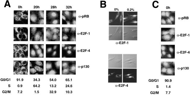
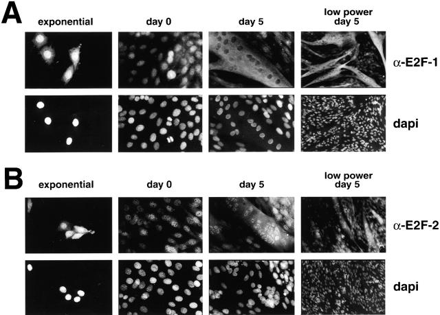
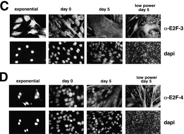
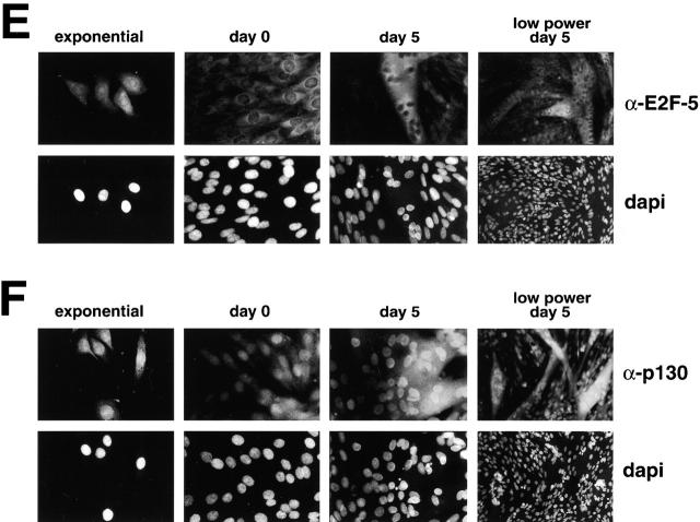
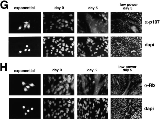
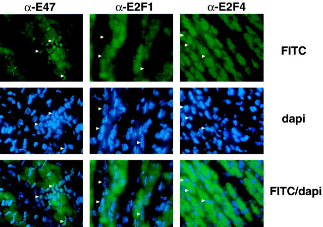
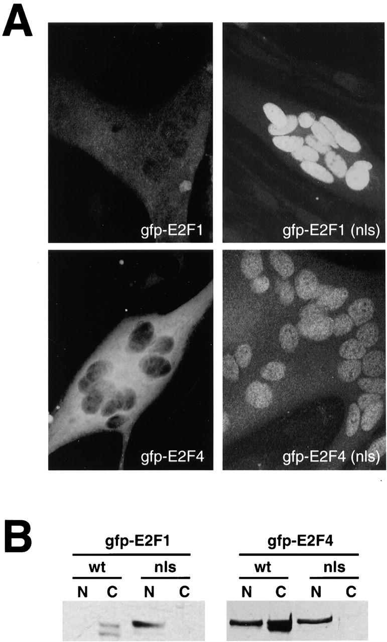
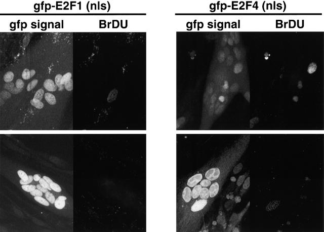
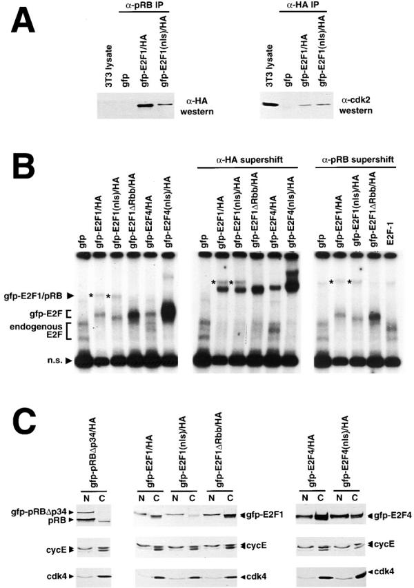
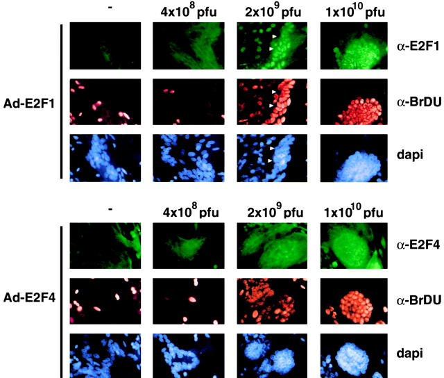
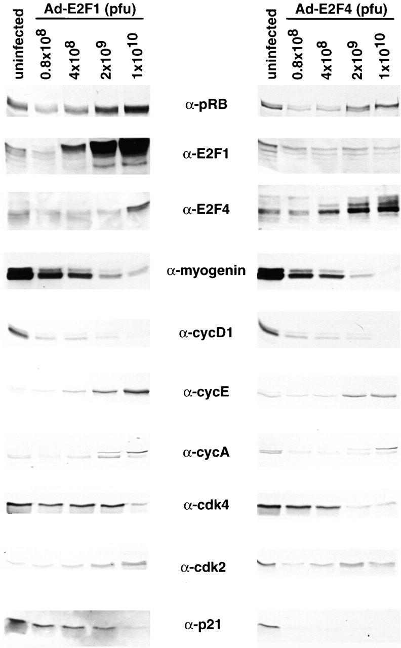
Maintenance of cells in a quiescent state after terminal differentiation occurs through a number of mechanisms that regulate the activity of the E2F family of transcription factors. We report here that changes in the subcellular compartmentalization of the E2F family proteins are required to prevent nuclei in terminally differentiated skeletal muscle from reentering S phase. In terminally differentiated L6 myotubes, E2F-1, E2F-3, and E2F-5 were primarily cytoplasmic, E2F-2 was nuclear, whereas E2F-4 became partitioned between both compartments. In these same cells, pRB family members, pRB, p107, and p130 were also nuclear. This compartmentalization of the E2F-1 and E2F-4 in differentiated muscle cells grown in vitro reflected their observed subcellular location in situ. We determined further that exogenous E2F-1 or E2F-4 expressed in myotubes at levels fourfold greater than endogenous proteins compartmentalized identically to their endogenous counterparts. Only when overexpressed at higher levels was inappropriate subcellular location for these proteins observed. At these levels, induction of the E2F-regulated genes, cyclins A and E, and suppression of factors associated with myogenesis, myogenin, and p21(Cip1) was observed. Only at these levels of E2F expression did nuclei in these terminally differentiated cells enter S phase. These data demonstrate that regulation of the subcellular compartmentalization of E2F-family members is required to maintain nuclei in a quiescent state in terminally differentiated cells.


Figures

Figure 1
Figure 1
E2F and pRB family members are compartmentalized during the cell cycle. (A) Rat-1 cells were growth-arrested in 0% serum for 40 h, followed by addition of 10% serum. Cells were trypsinized and collected for FACS analysis at 4-h intervals after serum addition and proportions of cells in G0/G1, S phase, and G2/M were determined. The subcellular localization of E2F and pRB members was determined by immunofluorescence as cells progress from G0 back into the cell cycle. Cells grown on coverslips, were removed at G0, S phase, G2/M, and the subsequent G1, fixed in −20°C methanol, and immunostained with antibodies against E2F-1, E2F-4, pRB, and p130. (B) The effect of a growth stimulus provided by low levels of growth factors on E2F subcellular localization was determined by arresting L6 myoblasts in either 0% serum or in 0.2% serum for 40 h. Cells grown on coverslips were removed, fixed in −20°C methanol and immunostained with antibodies against E2F-1 and E2F-4. Fixed cells are also visualized by light microscopy using Nomarski optics. (C) The effect of an ectopic growth stimulus on subcellular localization during the cell cycle was examined by arresting cells in the presence of inducible cyclin D1. Cells grown on coverslips in the absence of tetracycline (induced cyclin D1) were removed, fixed in −20°C methanol, and immunostained with antibodies against E2F-1, E2F-4, pRB, and p130.
Figure 2
Figure 2
E2F family members are differentially sequestered in the cytoplasm of terminally differentiated myotubes. The subcellular localization of E2F and pRB family members was examined by immunofluorescence as L6 myoblasts exit the cell cycle and terminally differentiate. Exponentially growing L6 cells were plated at 20% density and allowed to reach confluence, when serum was reduced to 2% (day 0) to induce differentiation to multinucleated postmitotic myotubes. Coverslips were removed from exponentially growing cultures, as well as at day 0, day 2, and day 5 after serum reduction. (A–E) Undifferentiated, day 0, day 2, and day 5 cells were immunostained with primary antibodies against E2F-1, E2F-2, E2F-3, E2F-4, and E2F-5 followed by FITC-conjugated secondary antibody. Nuclei are visualized by DAPI staining and the corresponding panels are presented below the appropriate FITC-labeled immunostained panel. (F–H) Undifferentiated, day 0, day 2, and day 5 cells were also immunostained with primary antibodies against pRB, p107, and p130 followed by FITC-conjugated secondary antibody. Undifferentiated cells are presented in the left-hand panels, and terminally differentiated multinucleated myotubes in the right-hand panels.
Figure 2
Figure 2
E2F family members are differentially sequestered in the cytoplasm of terminally differentiated myotubes. The subcellular localization of E2F and pRB family members was examined by immunofluorescence as L6 myoblasts exit the cell cycle and terminally differentiate. Exponentially growing L6 cells were plated at 20% density and allowed to reach confluence, when serum was reduced to 2% (day 0) to induce differentiation to multinucleated postmitotic myotubes. Coverslips were removed from exponentially growing cultures, as well as at day 0, day 2, and day 5 after serum reduction. (A–E) Undifferentiated, day 0, day 2, and day 5 cells were immunostained with primary antibodies against E2F-1, E2F-2, E2F-3, E2F-4, and E2F-5 followed by FITC-conjugated secondary antibody. Nuclei are visualized by DAPI staining and the corresponding panels are presented below the appropriate FITC-labeled immunostained panel. (F–H) Undifferentiated, day 0, day 2, and day 5 cells were also immunostained with primary antibodies against pRB, p107, and p130 followed by FITC-conjugated secondary antibody. Undifferentiated cells are presented in the left-hand panels, and terminally differentiated multinucleated myotubes in the right-hand panels.
Figure 3
Figure 3
E2F family members are localized in the cytoplasm of myotubes in the developing embryo. To determine whether cytoplasmic compartmentalization of the E2Fs occurs in vivo, sagittal sections of a murine day E18.5 embryo were immunostained against E2F-1, E2F-4, and as a control, the nuclear myogenic transcription factor, E47. Skeletal muscle sections adjacent to the developing ribs were probed with primary antibodies against E47, E2F-1, and E2F-4, followed by FITC-labeled secondary antibody. Nuclei are visualized by DAPI staining. FITC and DAPI signals were determined by fluorescent microscopy and are presented individually and as a combined FITC–DAPI image. Specific nuclei demonstrating nuclear immunostaining of E47 or nuclear exclusion of E2F-1 and E2F-4 are indicated by arrowheads.
Figure 4
Figure 4
Ectopic GFP-tagged E2F-1 and E2F-4 localize to the cytoplasm of myotubes and an ectopic NLS overcomes this compartmentalization. L6 cells were differentiated to multinucleated myotubes in 2% serum for 72 h. Myotubes were then infected with 5 × 10⁹ pfu of adenovirus expressing GFP–E2F-1 or GFP–E2F-4 fusion proteins, or GFP–E2F fusion proteins containing an ectopic SV40 LgT NLS. GFP signals were determined by fluorescent microscopy using the FITC stimulation wavelength. (A) Immunofluorescence of GFP-tagged E2F-1 (left side) or E2F-4 (right side) proteins without (top) or with (bottom) the SV40 NLS were visualized by fluorescence microscopy. (B) Localization of the GFP–E2F fusion proteins was also determined by Western analysis of fractionated lysates from infected cells. Nuclear and cytoplasmic lysates from terminally differentiated L6 myotube cultures infected with adenoviral vectors expressing GFP-tagged wild-type, or LgT NLS-tagged E2F-1 and E2F-4 were run on an SDS-PAGE gel and Western blotted with anti–E2F-1 (left panel) and anti–E2F-4 (right panel) polyclonal antibodies. Ectopic proteins are distinct from endogenous E2F due to the 25-kD GFP moiety.
Figure 5
Figure 5
Nuclear targeted GFP–E2F-1 and GFP–E2F-4 do not induce S phase in myotubes. L6 cells were differentiated to multinucleated myotubes in 2% serum for 72 h. Myotubes were then infected with 5 × 10⁹ pfu of adenovirus expressing GFP–E2F-1 (NLS) or GFP–E2F-4 (NLS) fusion proteins. 24 h after infection, BrDU labeling reagent was added and the cells were incubated for a further 48 h, at which point cells grown on coverslips were fixed, and nuclear and cytoplasmic lysates were prepared. Immunofluorescence of GFP-tagged E2F-1 (NLS) (left panel) or E2F-4 (NLS) (right panel) proteins was visualized by fluorescence microscopy using the FITC stimulation wavelength. S phase entry of GFP–E2F-expressing cells was determined by anti-BrdU immunohistochemistry using a Texas red–labeled anti–mouse secondary antibody.
Figure 6
Figure 6
GFP-tagged E2F molecules bind DNA and pRB family proteins but have altered transactivation properties. (A) To determine if endogenous pRB is capable of binding the GFP-tagged E2F-1 fusion proteins, 3T3 cells were infected with 1 × 10¹⁰ pfu of adenoviral vectors expressing GFP alone as well as GFP–E2F-1/HA and GFP–E2F-1 (NLS)/HA. 400 μg of whole cell lysate was immunoprecipitated with anti-pRB polyclonal antibody, and coimmunoprecipitating GFP-tagged E2F was determined by Western blotting with an anti-HA mAb. To determine whether the cyclin A binding domain is accessible, ectopic GFP-tagged E2F-1/HA and E2F-1 (NLS)/HA proteins, as well as a GFP control, were immunoprecipitated from adenoviral infected 3T3 lysates with monoclonal anti-HA antibody, and coimmunoprecipitating cdk2 protein was determined by anti-cdk2 Western blot (right panel). (B) Nuclear lysate from 3T3 cells infected with adenoviral vectors expressing either untagged E2F-1, or GFP-tagged wild-type, or NLS-tagged E2F-1 and E2F-4 was incubated at room temperature for 10 min with a 32P end-labeled double-stranded oligonucleotide probe containing a single E2F binding site, before resolving on a 4.5% nondenaturing acrylamide gel. The middle panel contains reactions in which 1 μl of monoclonal anti-HA was added for an additional 10 min, whereas the right panel contains reactions with anti-pRB monoclonal added. Endogenous E2F complexes are indicated, as are those containing GFP-tagged E2F. A larger complex containing pRB as well as ectopic GFP-tagged E2F-1 is indicated by an asterisk. Note that a mutant E2F molecule, which is deleted in the pRB-binding domain GFP–E2F-1ΔRbb/HA, does not form this pRB-containing complex. (C) 3T3 cells were infected with 1 × 10¹⁰ pfu of adenoviral vectors expressing either untagged E2F-1, or GFP-tagged wild-type, or NLS-tagged E2F-1 and E2F-4, as well as the pRB-binding mutant GFP–E2F-1ΔRbb/HA, and 72 h after infection, nuclear and cytoplasmic lysates were harvested and separated by SDS-PAGE. Levels of endogenous cyclin E protein were determined by Western blot. Exogenous GFP-tagged proteins are detected by an anti-HA monoclonal, and cdk4 is Western blotted as a control for protein loading.
Figure 7
Figure 7
Overexpression of ectopic E2F-1 and E2F-4 can overcome cytoplasmic sequestering and forces postmitotic myotube nuclei to enter S phase. L6 cells were differentiated to multinucleated myotubes in 2% serum for 48 h. Myotubes were then infected with the E2F-1 or E2F-4–expressing adenoviruses at either 0.8 × 10⁸, 4 × 10⁸, 2 × 10⁹, or 1 × 10¹⁰ pfu, and 48 h after infection DNA synthesis was determined by adding BrdU labeling reagent and incubating a further 24 h. Cells on coverslips were fixed and immunostained with mAb against BrdU and a Texas red–labeled anti–mouse secondary antibody, as well as polyclonal antibodies against E2F-1, or E2F-4, and FITC-labeled anti–rabbit secondary antibody. Nuclei were then stained with DAPI and each signal was captured by fluorescence microscopy. FITC, Texas red, and DAPI signals for myotubes infected with adenovirus expressing E2F-1 are shown in the top panel, whereas cells infected with E2F-4 adenovirus are shown in the bottom panel. Nuclei in Ad-E2F-1 infected cells (2 × 10⁹ pfu), which do not stain for BrdU or E2F-1, are indicated by arrowheads.
Figure 8
Figure 8
DNA synthesis caused by ectopic E2F-1 or E2F-4 is accompanied by changes in expression of cell cycle and differentiation-specific factors. L6 cells were differentiated to multinucleated myotubes in 2% serum for 48 h. Myotubes were then infected with the E2F-1 or E2F-4–expressing adenoviruses at either 4 × 10⁸, 2 × 10⁹, or 1 × 10¹⁰ pfu and 72 h after infection total lysates were isolated. 20 μg of lysate was run on an SDS-PAGE gel and Western blotted with antibodies against E2F-1, E2F-4, pRB, myogenin, p21Cip1, cdk2, cdk4, and cyclins D1, A, and E.

Similar articles

Cited by

References

    1. Aguzzi A., Kiess M., Rüedi D., Hamel P.A. Cyclins D1, D2 and D3 are expressed in distinct tissues during mouse embryogenesis. Transgenics. 1996;2:29–39.
    1. Arenzana-Seisdedos F., Turpin P., Rodriguez M., Thomas D., Hay R.T., Virelizier J.L., Dargemont C. Nuclear localization of I kappa B alpha promotes active transport of NF-kappa B from the nucleus to the cytoplasm. J. Cell Sci. 1997;110:369–378. - PubMed
    1. Bandara L.R., La Thangue N.B. Adenovirus E1a prevents the retinoblastoma gene product from complexing with a cellular transcription factor. Nature. 1991;351:494–497. - PubMed
    1. Beijersbergen R.L., Kerkhoven R.M., Zhu L., Carlee L., Voorhoeve P.M., Bernards R. E2F-4, a new member of the E2F gene family, has oncogenic activity and associates with p107 in vivo. Genes Dev. 1994;8:2680–2690. - PubMed
    1. Beijersbergen R.L., Carlee L., Kerkhoven R.M., Bernards R. Regulation of the retinoblastoma protein-related p107 by G1 cyclin complexes. Genes Dev. 1995;9:1340–1353. - PubMed

Publication types

MeSH terms