. 2000 Mar 20;148(6):1255-65.
doi: 10.1083/jcb.148.6.1255.

Kinesin superfamily protein 3 (KIF3) motor transports fodrin-associating vesicles important for neurite building

Affiliations

Kinesin superfamily protein 3 (KIF3) motor transports fodrin-associating vesicles important for neurite building

S Takeda et al. J Cell Biol. .

Abstract

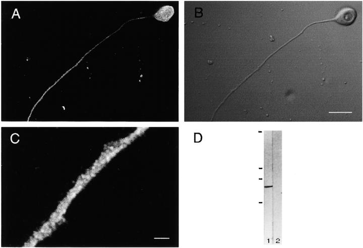
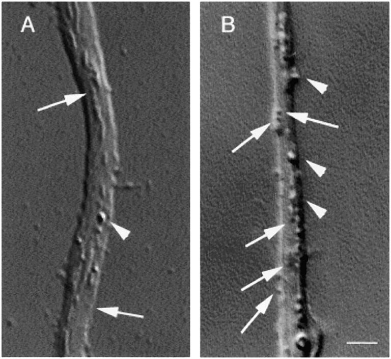
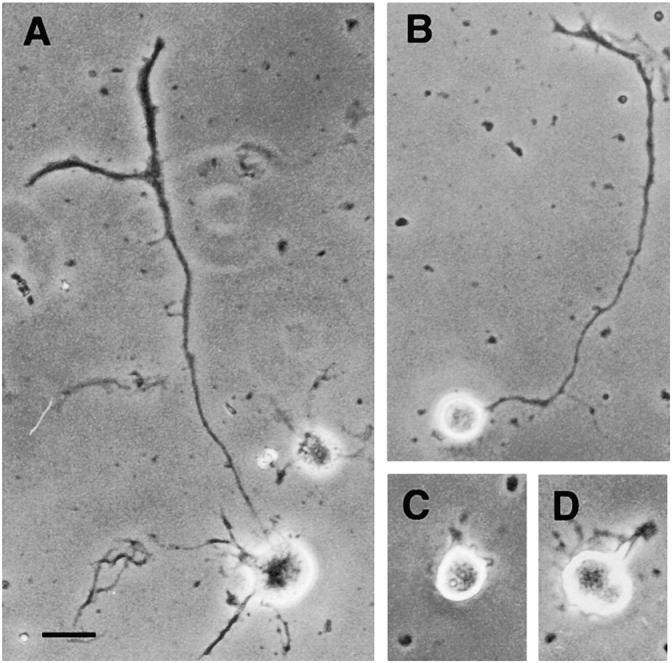
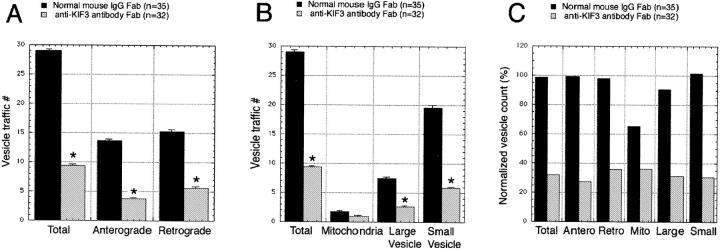
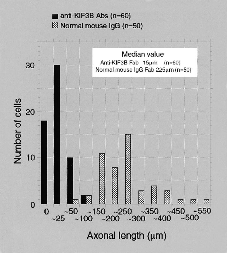
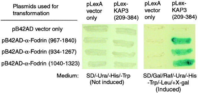
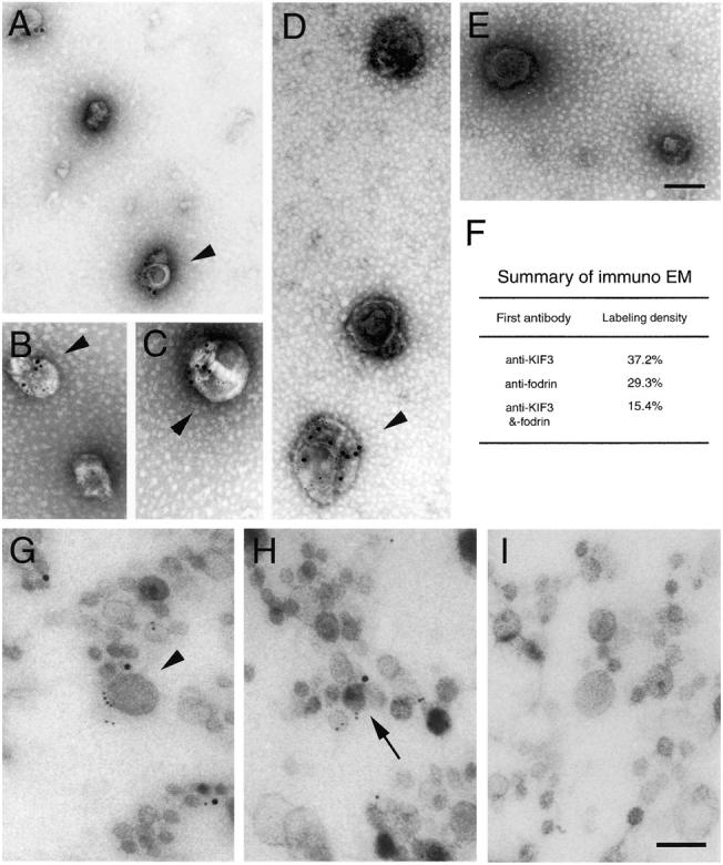
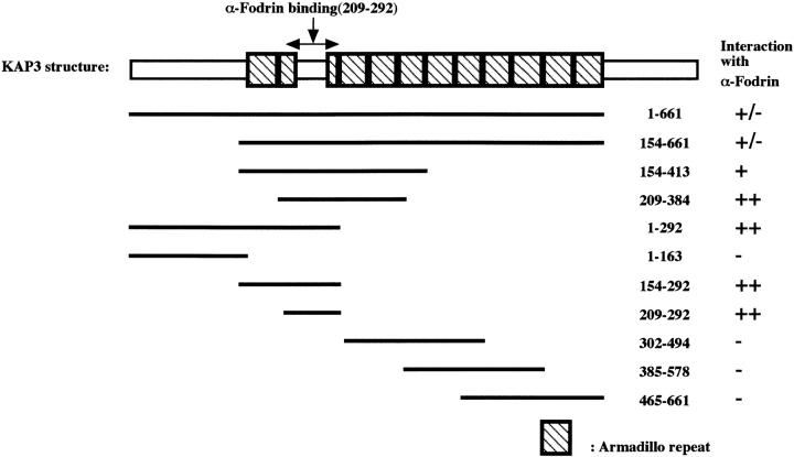
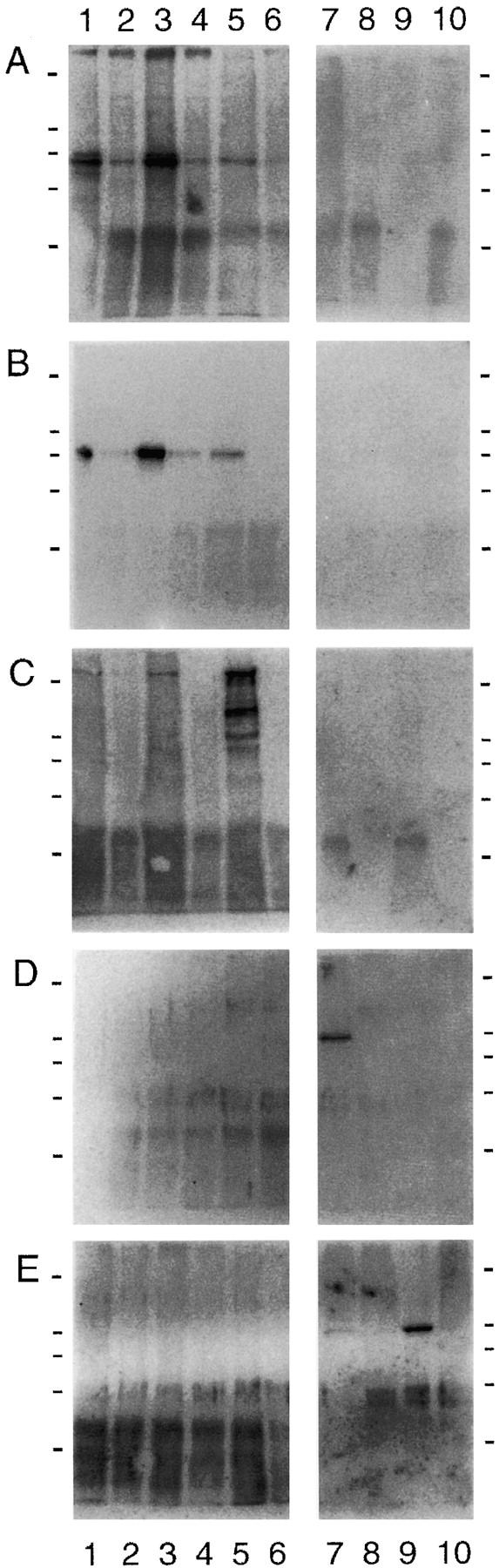
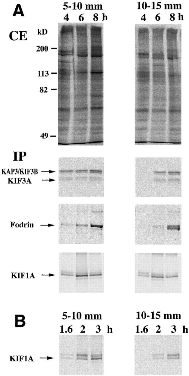
Kinesin superfamily proteins (KIFs) comprise several dozen molecular motor proteins. The KIF3 heterotrimer complex is one of the most abundantly and ubiquitously expressed KIFs in mammalian cells. To unveil the functions of KIF3, microinjection of function-blocking monovalent antibodies against KIF3 into cultured superior cervical ganglion (SCG) neurons was carried out. They significantly blocked fast axonal transport and brought about inhibition of neurite extension. A yeast two-hybrid binding assay revealed the association of fodrin with the KIF3 motor through KAP3. This was further confirmed by using vesicles collected from large bundles of axons (cauda equina), from which membranous vesicles could be prepared in pure preparations. Both immunoprecipitation and immunoelectron microscopy indicated the colocalization of fodrin and KIF3 on the same vesicles, the results reinforcing the evidence that the cargo of the KIF3 motor consists of fodrin-associating vesicles. In addition, pulse-labeling study implied partial comigration of both molecules as fast flow components. Taken together, the KIF3 motor is engaged in fast axonal transport that conveys membranous components important for neurite extension.


Figures

Figure 1
Figure 1
Characterization of anti-KIF3B antibody. A, Fluorescence immunocytochemical staining of cultured SCG neurons. Staining with anti-KIF3B antibody can be observed throughout the neuron, and is especially evident in axons. When viewed at higher magnification (C), a punctate staining pattern, suggestive of vesicular components, was also noted. B, DIC image of the same cell. D, Western blotting of mouse SCG extract probed with the anti-KIF3B antibody (lane 1). A single distinct band appears at a height corresponding to that of KIF3B, whereas no band is seen with preimmune serum (lane 2). Bars: (A and B) 20 μm; (C) 1 μm. Molecular weight bars in D (from top to bottom): 200, 116, 97, and 65 kD.
Figure 2
Figure 2
Examples of VEC–DIC images of SCG neuronal axons. SCG neurons grown on collagen type IV-treated coverslips were microinjected with either anti-KIF3B Fab or control normal mouse IgG Fab, and observed under VEC–DIC ∼3 h after microinjection. Microinjection of anti-KIF3B Fab brought about a decrease in the number of vesicles within the axons (A), whereas relatively ample vesicle traffic was seen in the axons microinjected with normal mouse IgG Fab (B). The arrowheads depict large vesicles; arrows indicate small vesicles. Bar, 1 μm.
Figure 4
Figure 4
Neurite sprouting was affected by microinjection of anti-KIF3B Fab. Either anti-KIF3B Fab or normal mouse IgG Fab was injected to the cultured SCG neurons before neurite sprouting. Approximately 12 h after injection, the cells were fixed and viewed by conventional fluorescence microscopy to identify the injected cells. Then, phase-contrast images of identified cells were taken. With normal mouse IgG Fab as the negative control, A and B, the treated cells extended axons in an ordinary fashion with ramification. On the contrary, microinjection of anti-KIF3B Fab, C and D, had a drastic effect, and most of the SCG neurons did not extend any neurites at all or exhibited only miniature neurites. Bar, 20 μm.
Figure 3
Figure 3
Comparison of vesicle traffic before and after microinjection of Fabs. A, Bidirectional effect of anti-KIF3B Fab microinjection. Both antero- and retrograde traffic were inhibited by the microinjection of the Fab fragments, although the degree of inhibition of the anterograde traffic was stronger. B, Each category of vesicles was counted by the number passing through a unit square (see Observation and Data Collection in Material and Methods, n/μm2/5 min) of axonal cross-section. A significant decrease in vesicle traffic was recorded, except in mitochondria. Five comparisons yielded significant P values (P < 0.05; *). Bars indicate SEM. C, Normalized vesicle traffic calibrated with that of uninjected cells. For both KIF3B Fab- and normal mouse IgG Fab-injected cells, the number of vesicles passing through a unitary axon transverse section was divided by that in uninjected cells. Note that the injection process, per se, did not have any deleterious effect on the vesicle traffic (the normal mouse IgG Fab group exhibited nearly 100% vesicle traffic in almost all subcategories, compared to the uninjected group).
Figure 5
Figure 5
Graphic summary for the axonal length in each experimental group. Distribution of neurite length after microinjection of anti-KIF3B Fab (black) and normal mouse IgG Fab (hatched). Most of the SCG neurons microinjected with anti-KIF3B Fab (n = 60) had short axons, as represented in Fig. 4C and Fig. D, whereas the SCG neurons injected with normal mouse IgG Fab extended their axon normally. The median values for each experimental group show that anti-KIF3B Fab dramatically inhibited (15 μm) axonal outgrowth, compared to normal mouse IgG Fab (225 μm).
Figure 6
Figure 6
Yeast two-hybrid assay of KAP3 with vesicle-associated proteins. After a yeast two-hybrid system screening, the interaction of α-fodrin with KAP3 was confirmed reproducibly by retransformation. Yeasts containing each combination of pLexA (no insert or KAP3) and pBD42AD constructs (no insert, α-fodrin of various lengths, aa 967–1840, 934–1267, and 1040–1323) grew on the SD/−His/−Trp/−Ura plate containing glucose (left), although those with only α-fodrin survived (right) on the selection plate (SD/−His/−Trp/−Ura/−Leu containing galactose/raffinose/X-gal/BU salts), showing blue colonies.
Figure 9
Figure 9
Immuno-colocalization of KIF3 and fodrin on cauda equina vesicles. A–D, Negatively stained cauda equina vesicles containing both KIF3B and fodrin. Vesicles ranging from 50–200 nm in diameter were double-labeled (arrowheads). 10-nm gold particles correspond to the localization of KIF3B and 5-nm gold particles depict αII-fodrin localization. E, Negative control probed with preimmune serum and normal mouse IgG, followed by probing with the auroprobe. No labeling was observed in this panel, but total nonspecific labeling was 4.0%. F, Diagram summarizing the frequency of labeling for KIF3B, fodrin, or both. About 37% of all vesicles contained the KIF3B labeling. G and H, Ultrathin sections of cauda equina vesicles labeled with both anti-KIF3 and αII-fodrin antibody. Compared to the former technique, smaller vesicles, <50 nm in diameter, could be visualized to contain the labeling (H, arrow). I, Negative control incubated with preimmune serum and normal mouse IgG, followed by the reaction with auroprobe. Bars, 100 nm.
Figure 7
Figure 7
Schematic representation of the functional domain of KAP3 and the α-fodrin molecule obtained by the yeast two-hybrid system. This scheme depicts the entire amino acid sequence of the KAP3 molecule (1–793). We constructed bait vectors of various lengths. The minimal required domain for interaction with fodrin consisted of aa 209–292, as indicated. Determination of the binding affinity using liquid yeast cultures was also performed, and the results are summarized at the right of the figure. The minimal required domain (209–291) showed strongest binding affinity (indicated by ++).
Figure 8
Figure 8
Immunoprecipita-tion of the vesicle fraction of cauda equina by anti-KIF3B and KAP3 antibodies. Cauda equina vesicles were immunoprecipitated with either anti-KIF3B (lane 1), anti-KAP3 (lane 3), anti–αII-fodrin (lane 5), anti-KIF5A (lane 7), or anti-KIF5B (lane 9) beads. To check the interaction, these immunoprecipitation products were probed with anti-KIF3B (A), anti-KAP3 (B), anti–αII-fodrin (C), anti-KIF5A (D), and anti-KIF5B (E) antibodies. Equal amounts of beads were loaded onto the SDS gel and analyzed by Western blotting under the same condition. Note that the interaction between the KIF3 complex and fodrin could be bilaterally confirmed. Furthermore, fodrin does not associate with KIF5A or KIF5B. In this experiment, immunoprecipitated cauda equina vesicles were solubilized with 1% Triton X-100 for 1 h on ice. The solubilization did not destroy the interaction between fodrin and the KIF3 motor. Lanes 2, 4, 6, 8, and 10 are negative controls immunoprecipitated with the beads treated with the preimmune serum (2, 4, 8, and 10) or normal mouse IgG (6) instead of the specific immunoglobulin for each antigens. Molecular weight bars (from top to bottom): 200, 116, 97, 65, and 45 kD.
Figure 10
Figure 10
Pulse-labeling study revealing the velocity of KIF3, KIF1A, and fodrin in the rat optic nerve. A, [35S]methionine was injected into the vitreous. 4, 6, and 8 h after administration, the optic nerve and tract were extirpated en bloc and further cut into consecutive 5-mm segments. The second (5–10 mm) and third (10–15 mm) segments were used for the analyses. Note that KIF3 and fodrin bands begin to appear 6 h after treatment. B, Since the reported velocity of KIF1A is much higher than that of KIF3, we killed the animals earlier for determination of the former. In this case, the time of appearance of the KIF1A bands shows good agreement with previous in vitro data, confirming the appropriateness of this system for roughly estimating the velocity of axonal transport. CE, Crude extract; IP, immunoprecipitation.

Similar articles

Cited by

References

    1. Aizawa H., Sekine Y., Takemura R., Zhang Z., Nangaku M., Hirokawa N. Kinesin family in murine central nervous system. J. Cell Biol. 1992;119:1287–1296. - PMC - PubMed
    1. Ausubel, F.M., R. Brent, R.E. Kingston, D.D. Moore, J.G. Seidman, J.A. Smith, and K. Struhl. 1997. Current Protocols in Molecular Biology. John Wiley & Sons, Inc. New York. 13.6.2–13.6.5.
    1. Bamburg J.R., Bray D., Chapman K. Assembly of microtubules at the tip of growing axons. Nature. 1986;321:788–790. - PubMed
    1. Beech P.L., Pagh-Roehl K., Noda Y., Hirokawa N., Burnside B., Rosenbaum J.L. Localization of kinesin superfamily proteins to the connecting cilium of fish photoreceptors. J. Cell Sci. 1996;109:889–897. - PubMed
    1. Brady S.T. A novel brain ATPase with properties expected for the fast axonal transport motor. Nature. 1985;317:73–75. - PubMed

Publication types

MeSH terms