. 2000 Mar 20;148(6):1267-81.
doi: 10.1083/jcb.148.6.1267.

beta-arrestin-dependent endocytosis of proteinase-activated receptor 2 is required for intracellular targeting of activated ERK1/2

Affiliations

beta-arrestin-dependent endocytosis of proteinase-activated receptor 2 is required for intracellular targeting of activated ERK1/2

K A DeFea et al. J Cell Biol. .

Abstract

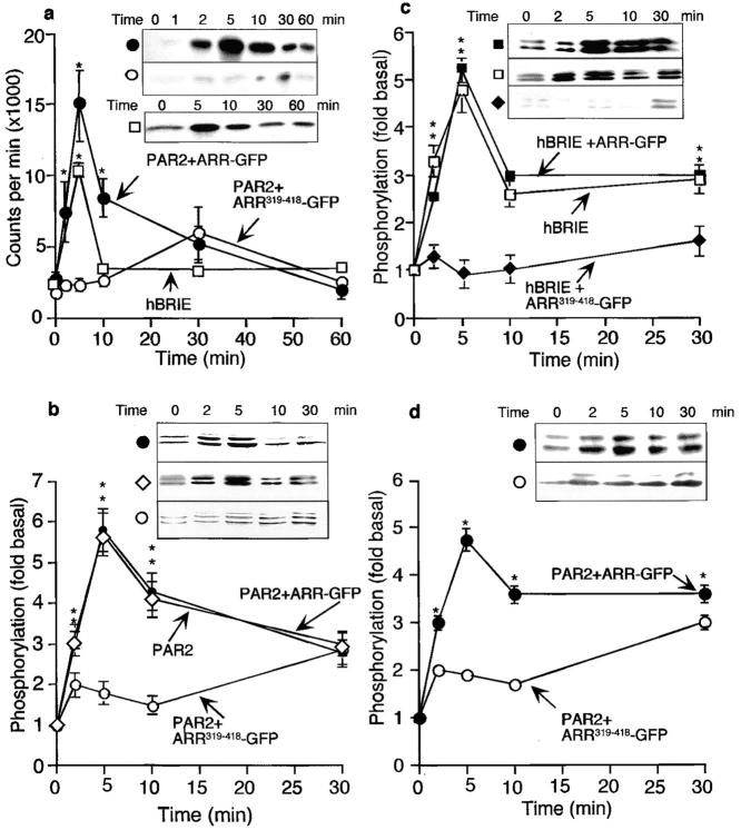
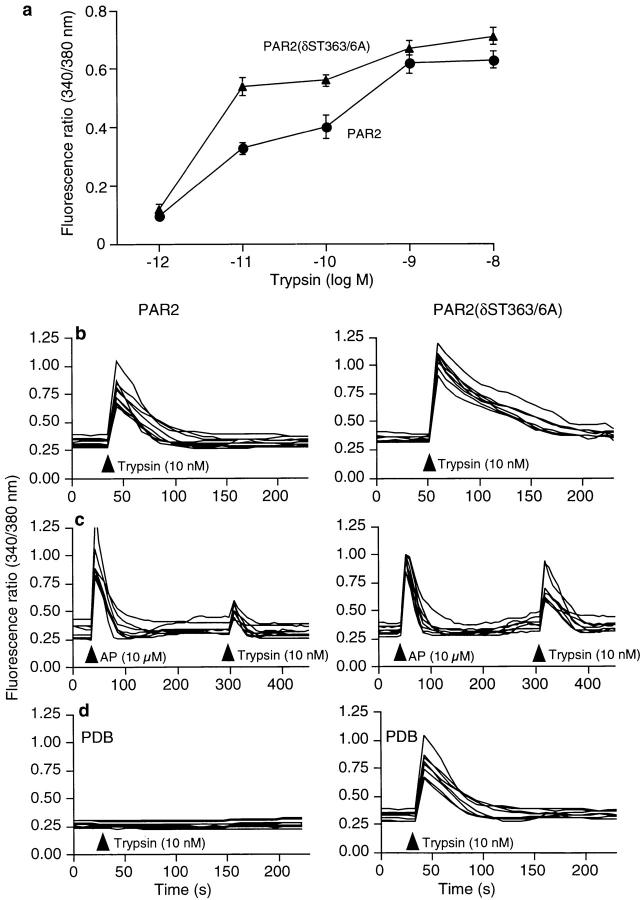
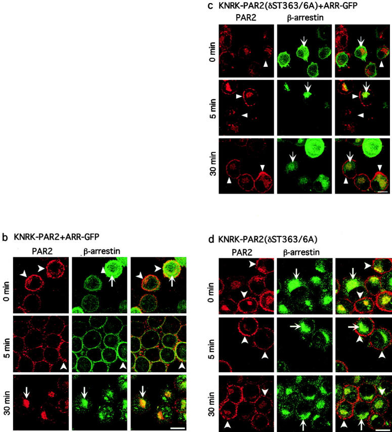
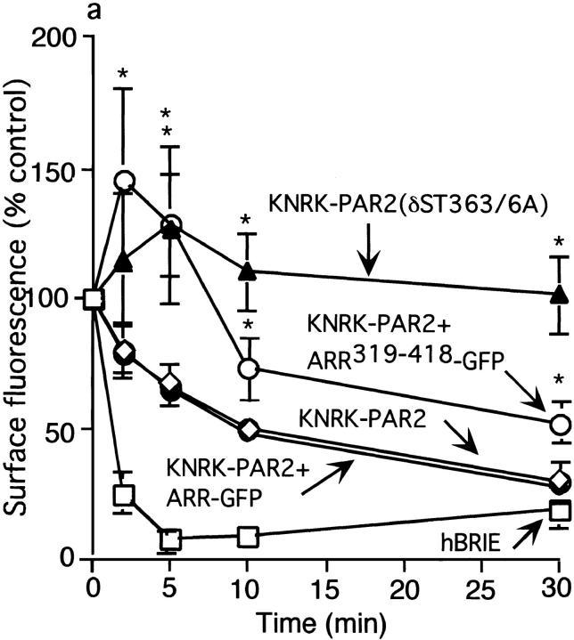
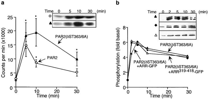
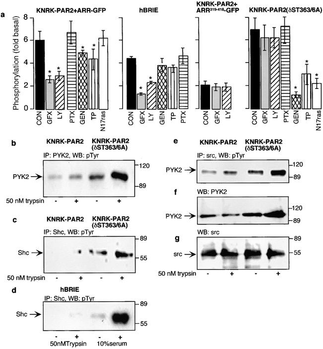
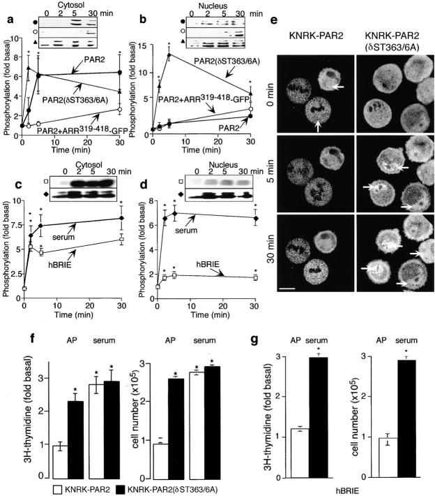
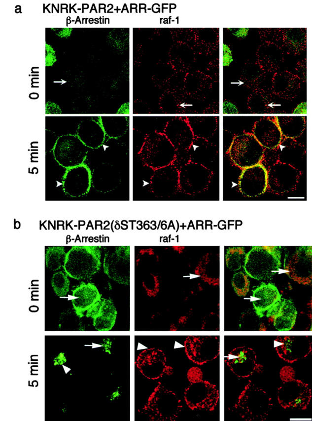
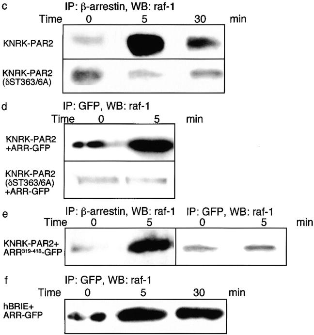
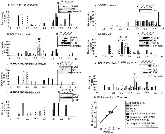
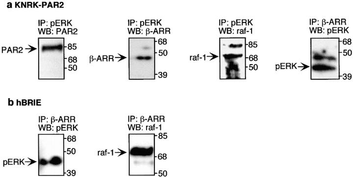
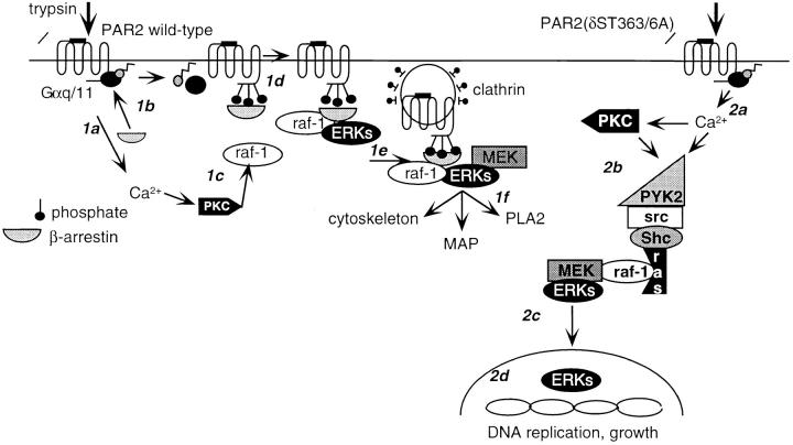
Recently, a requirement for beta-arrestin-mediated endocytosis in the activation of extracellular signal-regulated kinases 1 and 2 (ERK1/2) by several G protein-coupled receptors (GPCRs) has been proposed. However, the importance of this requirement for function of ERK1/2 is unknown. We report that agonists of Galphaq-coupled proteinase-activated receptor 2 (PAR2) stimulate formation of a multiprotein signaling complex, as detected by gel filtration, immunoprecipitation and immunofluorescence. The complex, which contains internalized receptor, beta-arrestin, raf-1, and activated ERK, is required for ERK1/2 activation. However, ERK1/2 activity is retained in the cytosol and neither translocates to the nucleus nor causes proliferation. In contrast, a mutant PAR2 (PAR2deltaST363/6A), which is unable to interact with beta-arrestin and, thus, does not desensitize or internalize, activates ERK1/2 by a distinct pathway, and fails to promote both complex formation and cytosolic retention of the activated ERK1/2. Whereas wild-type PAR2 activates ERK1/2 by a PKC-dependent and probably a ras-independent pathway, PAR2(deltaST363/6A) appears to activate ERK1/2 by a ras-dependent pathway, resulting in increased cell proliferation. Thus, formation of a signaling complex comprising PAR2, beta-arrestin, raf-1, and activated ERK1/2 might ensure appropriate subcellular localization of PAR2-mediated ERK activity, and thereby determine the mitogenic potential of receptor agonists.


Figures

Figure 1
Figure 1
PAR2-mediated activation of ERK1/2. (a) KNRK-PAR2+ARR-GFP cells (•) and KNRK-PAR2+ARR319-418-GFP cells (○) and hBRIE cells (□) were incubated with 50 nM trypsin for 0–60 min at 37°C, and ERK activity was measured using the MBP assay. (b–d) Western blots using antibodies to pERK1/2. (b) KNRK-PAR2+ARR-GFP cells (•), KNRK-PAR2 cells (⋄), and KNRK-PAR2+ARR319-418-GFP cells (○) were incubated with 50 nM trypsin. (c) hBRIE cells (□), hBRIE+ARR-GFP (⋄), and hBRIE+ARR319-418-GFP (♦) were incubated with 50 nM trypsin for 0–30 min at 37°C. (d) KNRK-PAR2+ARR-GFP cells (•) and KNRK-PAR2+ARR319-418-GFP cells (○) were incubated 50 μM AP for 0–30 min at 37°C. *P < 0.05 compared with cells expressing PAR2 alone or PAR2 plus ARR-GFP cells, n = 4.
Figure 2
Figure 2
PAR2-mediated Ca2+ mobilization in KNRK-PAR2 cells and KNRK-PAR2δ(ST363/6A) cells. (a) Concentration-response analysis for KNRK-PAR2 (•) and KNRK-PAR2 (δST363/6A) (▴) cells. (b–d) Each line shows [Ca2+]i for individual KNRK-PAR2 cells (left) and KNRK-PAR2(δST363/6A) cells (right). (b) Note that the response to trypsin is prolonged in KNRK-PAR2(δST363/6A) cells. (c) Homologous desensitization in cells pretreated with 10 μM AP for 5 min before addition of 10 nM trypsin. (d) Heterologous desensitization in cells pretreated with 1 μM PDB for 20 min before addition of 10 nM trypsin. Note that KNRK-PAR2(δST363/6A) cells are resistant to desensitization.
Figure 3
Figure 3
Agonist-induced PAR2 internalization. (a) Kinetics of PAR2 endocytosis in KNRK-PAR2+ARR-GFP cells (•), KNRK-PAR2 cells (⋄) KNRK-PAR2+ ARR319-418-GFP cells (○), KNRK-PAR2(δST363/6A) cells (▴), and hBRIE cells (□). Cells were incubated with 50 μM AP for 0–30 min at 37°C and endocytosis was determined by measuring surface Flag immunoreactivity by flow cytometry. *P < 0.05 compared with cells expressing PAR2 alone or PAR2 plus ARR-GFP cells, n = 3. (b–d) Localization of PAR2 and β-arrestin by immunofluorescence and confocal microscopy. KNRK-PAR2+ARR-GFP cells (b), KNRK-PAR2(δST363/6A)+ ARR-GFP cells (c), or KNRK-PAR2(δST363/6A) (d) were incubated with 50 nM trypsin for 0–30 min at 37°C. PAR2 was localized by immunofluorescence and β-arrestin was detected using GFP (b and c) or by immunofluorescence (d). The same cells are shown in each row and the images in the right panel are formed by superimposition of the images from the other two panels in the same row. Representative of two experiments. In KNRK-PAR2+ARR-GFP cells, note redistribution of β-arrestin to the plasma membrane at 5 min (arrowheads) and to endosomes at 30 min (arrows), where it colocalizes with PAR2. In KNRK-PAR2(δST363/6A)+ARR-GFP cells and in KNRK-PAR2(δST363/6A), note that PAR2 remains at the plasma membrane (arrowheads) and β-arrestin remains in the cytosol (arrows) with no colocalization. Bar, 10 μm.
Figure 3
Figure 3
Agonist-induced PAR2 internalization. (a) Kinetics of PAR2 endocytosis in KNRK-PAR2+ARR-GFP cells (•), KNRK-PAR2 cells (⋄) KNRK-PAR2+ ARR319-418-GFP cells (○), KNRK-PAR2(δST363/6A) cells (▴), and hBRIE cells (□). Cells were incubated with 50 μM AP for 0–30 min at 37°C and endocytosis was determined by measuring surface Flag immunoreactivity by flow cytometry. *P < 0.05 compared with cells expressing PAR2 alone or PAR2 plus ARR-GFP cells, n = 3. (b–d) Localization of PAR2 and β-arrestin by immunofluorescence and confocal microscopy. KNRK-PAR2+ARR-GFP cells (b), KNRK-PAR2(δST363/6A)+ ARR-GFP cells (c), or KNRK-PAR2(δST363/6A) (d) were incubated with 50 nM trypsin for 0–30 min at 37°C. PAR2 was localized by immunofluorescence and β-arrestin was detected using GFP (b and c) or by immunofluorescence (d). The same cells are shown in each row and the images in the right panel are formed by superimposition of the images from the other two panels in the same row. Representative of two experiments. In KNRK-PAR2+ARR-GFP cells, note redistribution of β-arrestin to the plasma membrane at 5 min (arrowheads) and to endosomes at 30 min (arrows), where it colocalizes with PAR2. In KNRK-PAR2(δST363/6A)+ARR-GFP cells and in KNRK-PAR2(δST363/6A), note that PAR2 remains at the plasma membrane (arrowheads) and β-arrestin remains in the cytosol (arrows) with no colocalization. Bar, 10 μm.
Figure 4
Figure 4
(a) PAR2-mediated ERK1/2 activity. KNRK-PAR2 (⋄) and KNRK-PAR2(δST363/6A) (▴) cells were treated with 50 nM trypsin for 0–30 min, and ERK activity was measured using the MBP assay. (b) Activation of ERK1/2. KNRK-PAR2(δST363/6A) (▴), KNRK-PAR2 (δST363/6A)+ARR-GFP (▵), and KNRK-PAR2 (δST363/6A)+ARR319-418-GFP (♦) cells were treated with 50 nM trypsin for 0–30 min and phosphorylation was assessed using antibodies to pERK1/2. *P < 0.05 compared with KNRK-PAR2 cells, n = 3.
Figure 5
Figure 5
Mechanism of PAR2-mediated activation of ERK1/2. (a) KNRK-PAR2, hBRIE, KNRK-PAR2+ARR319-418, and KNRK-PAR2(δST363/6A) cells were untreated (control, con), or incubated with 100 nM GF109203X (GFX), 20 nM LY379196 (LY), 100 ng/ml PTX, 10 μM genistein (GEN), 20 μM tyrphostin 25 (TP), or were cotransfected with N17ras. Cells were incubated with 50 nM trypsin for 5 min. *P < 0.05 compared with untreated cells, n = 4. (b–f) Analysis of KNRK-PAR2 and KNRK-PAR2(δST363/6A) cells by immunoprecipitation (IP) and Western blotting (WB). Cells were incubated with 50 nM trypsin for 5 min. Extracts were immunoprecipitated with antibodies to PYK2 (b), Shc (c and d), and src (e–g). Blots were probed for phosphotyrosine (b–e), PYK2 (f), and src (g).
Figure 6
Figure 6
Nuclear translocation of ERK1/2. (a–d) Subcellular fractionation of activated ERK1/2. KNRK-PAR2+ARR-GFP cells (•), KNRK-PAR2+ ARR319-418-GFP cells (○), or PAR2(δST363/6A) cells (s) were incubated with 50 nM trypsin for 0–30 min at 37°C, and pERK was determined in the cytosolic (a) and nuclear (b) fractions (n = 3). hBRIE cells were incubated with 50 nM trypsin (□) or 10% serum (♦), and pERK was determined in the cytosolic (c) and nuclear (d) fractions. (e) KNRK-PAR2 and KNRK-PAR2(δST363/6A) cells, transiently transfected with ERK2-GFP, were incubated with 50 nM trypsin at 37°C, and translocation of ERK2-GFP was observed by confocal imaging. Representative of eight experiments. In KNRK-PAR2 cells, note that ERK2-GFP remains cytosolic but, in KNRK-PAR2(δST363/6A) cells, it redistributes to the nucleus. (f and g) Proliferative responses to AP and serum. KNRK-PAR2 (f, □) and KNRK-PAR2(δST363/6A) cells (f, ▪) or hBRIE cells (g) were incubated with 50 μM AP or 20% FCS for 24 h, and incorporation of [3H]thymidine and cell number were measured. *P < 0.05 compared with untreated cells or KNRK-PAR2 cells, n = 3.
Figure 7
Figure 7
Trypsin induced association of β-arrestin and raf-1. (a and b) Localization of β-arrestin and raf-1 by immunofluorescence and confocal microscopy. KNRK-PAR2+ARR-GFP cells (a) or KNRK-PAR2(δST363/6A)+ ARR-GFP cells (b) were incubated with 50 nM trypsin for 0 or 5 min at 37°C. β-Arrestin was localized using GFP and raf-1 was localized by immunofluorescence. The same cells are shown in each row and the images in the right panel are formed by superimposition of images from the other two panels in the same row. Representative of two experiments. In KNRK-PAR2+ARR-GFP cells, note the redistribution of β-arrestin and raf-1 from the cytosol at 0 min (arrows) to the plasma membrane at 5 min (arrowheads), where they colocalize. In KNRK-PAR2 (δST363/6A)+ARR-GFP cells, note that β-arrestins remain in the cytosol (arrows) and raf-1 redistributes to the plasma membrane at 5 min (arrowheads). (c–f) Coimmunoprecipitation of raf-1 and β-arrestin. Cells were incubated with 50 nM trypsin for 0–30 min at 37°C, lysed, immunoprecipitated (IP) using antibodies to GFP or β-arrestin-1/2, and analyzed by Western blotting (WB) with a raf-1 antibody. (c) In KNRK-PAR2 cells, but not in KNRK-PAR2(δST363/6A) cells, β-arrestin and raf-1 coprecipitated. (d) Similarly, in KNRK-PAR2+ARR-GFP cells but not KNRK-PAR2(δST363/6A)+ARR-GFP cells ARR-GFP and raf-1 coprecipitated with antibodies to GFP. (e) In KNRK-PAR2+ARR319-418-GFP cells, endogenous β-arrestin and raf-1 coprecipitated, but ARR319-418 and raf-1 did not coprecipitate. (f) In hBRIE+ARR-GFP cells, ARR-GFP and raf-1 coprecipitated. Bars, 10 μm.
Figure 7
Figure 7
Trypsin induced association of β-arrestin and raf-1. (a and b) Localization of β-arrestin and raf-1 by immunofluorescence and confocal microscopy. KNRK-PAR2+ARR-GFP cells (a) or KNRK-PAR2(δST363/6A)+ ARR-GFP cells (b) were incubated with 50 nM trypsin for 0 or 5 min at 37°C. β-Arrestin was localized using GFP and raf-1 was localized by immunofluorescence. The same cells are shown in each row and the images in the right panel are formed by superimposition of images from the other two panels in the same row. Representative of two experiments. In KNRK-PAR2+ARR-GFP cells, note the redistribution of β-arrestin and raf-1 from the cytosol at 0 min (arrows) to the plasma membrane at 5 min (arrowheads), where they colocalize. In KNRK-PAR2 (δST363/6A)+ARR-GFP cells, note that β-arrestins remain in the cytosol (arrows) and raf-1 redistributes to the plasma membrane at 5 min (arrowheads). (c–f) Coimmunoprecipitation of raf-1 and β-arrestin. Cells were incubated with 50 nM trypsin for 0–30 min at 37°C, lysed, immunoprecipitated (IP) using antibodies to GFP or β-arrestin-1/2, and analyzed by Western blotting (WB) with a raf-1 antibody. (c) In KNRK-PAR2 cells, but not in KNRK-PAR2(δST363/6A) cells, β-arrestin and raf-1 coprecipitated. (d) Similarly, in KNRK-PAR2+ARR-GFP cells but not KNRK-PAR2(δST363/6A)+ARR-GFP cells ARR-GFP and raf-1 coprecipitated with antibodies to GFP. (e) In KNRK-PAR2+ARR319-418-GFP cells, endogenous β-arrestin and raf-1 coprecipitated, but ARR319-418 and raf-1 did not coprecipitate. (f) In hBRIE+ARR-GFP cells, ARR-GFP and raf-1 coprecipitated. Bars, 10 μm.
Figure 8
Figure 8
Gel filtration analysis of an ERK signaling complex. KNRK-PAR2 (a and b), KNRK-PAR2(δST363/6A) cells (c and d), or hBRIE cells (e and f), or KNRK-PAR2+ARR319-418-GFP cells (g) were untreated (a, c, and e) or incubated with 50 μM AP (b, d, f, and g) for 5 min. Cell lysates were fractionated on a S300 Sephacryl column. The presence of pERK, raf-1, β-arrestin-1, and PAR2 in each fraction was determined by Western blotting (inset). PAR2 was detected using HA.11 antibody in KNRK cells and 2N antibody in hBRIE cells. Results are expressed as a percentage of the total protein for each partition coefficient (σ) (n = 3). The bracketed columns represent regions where proteins coeluted. Representative Western blots are shown of fractions containing the complex in KNRK-PAR2 cells (★, σ = 0.34), hBRIE cells (⋆, σ = 0.31–34), and in KNRK-PAR2+ARR319-418-GFP (*, σ = 0.45). (h) The Stoke's radii of the four molecular mass standards and the complex in KNRK-PAR2 cells (★, ∼6.2), hBRIE cells (⋆, ∼6.2–6.6), and KNRK-PAR2+ARR319-418-GFP (*, ∼5 nm) are graphed as a function of the error function complement of σ.
Figure 9
Figure 9
Coprecipitation of components of the β-arrestin–containing signaling complex. (a) Fractions from the gel filtration columns of AP-stimulated KNRK-PAR2 cells at partition coefficients 0.31–0.34 were pooled, concentrated, and immunoprecipitated with pERK or β-arrestin-1/2 antibodies. Western blots were probed for PAR2 using the HA.11 antibody, β-arrestin-1, raf-1, and pERK. (b) Fractions from the gel filtration columns of AP-stimulated hBRIE cells at partition coefficients 0.31–0.34 were similarly processed, immunoprecipitated with a β-arrestin-1/2 antibody, and blotted with antibodies to pERK and raf-1.
Figure 10
Figure 10
Model for β-arrestin–dependent and –independent activation of ERK1/2 by PAR2. (1a) Trypsin cleavage and activation of wild-type PAR2 leads to transient Ca2+ mobilization and activation of PKC. (1b) PKC phosphorylates and activates raf-1. (1c) PAR2 binds β-arrestin and desensitizes. (1d) Raf-1 and ERK1/2 associate with β-arrestin–bound receptor and (1e) the complex associates with the clathrin-coated pit and ERK becomes activated. (1f) Cytosolic, active ERK1/2 phosphorylate cytoskeletal proteins, microtubule-associated proteins (MAPs), and phospholipase A2 (PLA2). (2a) Trypsin cleavage and activation of PAR2(δST363/6A) leads to sustained Ca2+ mobilization and transactivation of EGFR. (2b) Tyrosine phosphorylation of kinases such as PYK2 occur, activating the classic ras-dependent ERK1/2 pathway. (2c) ERK1/2 translocate to the nucleus. (2d) ERK1/2 phosphorylate and activate nuclear proteins, leading to DNA replication and cell growth.

Similar articles

Cited by

References

    1. Alblas J., van Etten I., Moolenaar W.H. Truncated, desensitization-defective neurokinin receptors mediate sustained MAP kinase activation, cell growth and transformation by a Ras-independent mechanism. EMBO (Eur. Mol. Biol. Organ.) J. 1996;15:3351–3360. - PMC - PubMed
    1. Böhm S.K., Khitin L., Smeekens S.P., Grady E.F., Payan D.G., Bunnett N.W. Identification of potential tyrosine-containing endocytic motifs in the carboxyl-tail and seventh transmembrane domain of the neurokinin 1 receptor. J. Biol. Chem. 1997;272:2363–2372. - PubMed
    1. Böhm S.K., Khitin L.M., Grady E.F., Aponte G., Payan D.G., Bunnett N.W. Mechanisms of desensitization and resensitization of proteinase-activated receptor-2. J. Biol. Chem. 1996;271:22003–22016. - PubMed
    1. Bornfeldt K.E., Campbell J.S., Koyama H., Argast G.M., Leslie C.C., Raines E.W., Krebs E.G., Ross R. The mitogen-activated protein kinase pathway can mediate growth inhibition and proliferation in smooth muscle cells. Dependence on the availability of downstream targets. J. Clin. Invest. 1997;100:875–885. - PMC - PubMed
    1. Brunet A., Roux D., Lenormand P., Dowd S., Keyse S., Pouysségur J. Nuclear translocation of p42/p44 mitogen-activated protein kinase is required for growth factor-induced gene expression and cell cycle entry. EMBO (Eur. Mol. Biol. Organ.) J. 1999;18:664–674. - PMC - PubMed

Publication types

MeSH terms