The tissue plasminogen activator (tPA)/plasmin extracellular proteolytic system regulates seizure-induced hippocampal mossy fiber outgrowth through a proteoglycan substrate
- PMID: 10725341
- PMCID: PMC2174310
- DOI: 10.1083/jcb.148.6.1295
The tissue plasminogen activator (tPA)/plasmin extracellular proteolytic system regulates seizure-induced hippocampal mossy fiber outgrowth through a proteoglycan substrate
Abstract
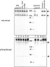
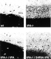
Short seizure episodes are associated with remodeling of neuronal connections. One region where such reorganization occurs is the hippocampus, and in particular, the mossy fiber pathway. Using genetic and pharmacological approaches, we show here a critical role in vivo for tissue plasminogen activator (tPA), an extracellular protease that converts plasminogen to plasmin, to induce mossy fiber sprouting. We identify DSD-1-PG/phosphacan, an extracellular matrix component associated with neurite reorganization, as a physiological target of plasmin. Mice lacking tPA displayed decreased mossy fiber outgrowth and an aberrant band at the border of the supragranular region of the dentate gyrus that coincides with the deposition of unprocessed DSD-1-PG/phosphacan and excessive Timm-positive, mossy fiber termini. Plasminogen-deficient mice also exhibit the laminar band and DSD- 1-PG/phosphacan deposition, but mossy fiber outgrowth through the supragranular region is normal. These results demonstrate that tPA functions acutely, both through and independently of plasmin, to mediate mossy fiber reorganization.
Figures


Similar articles
-
Phospholipase D1-promoted release of tissue plasminogen activator facilitates neurite outgrowth.J Neurosci. 2005 Feb 16;25(7):1797-805. doi: 10.1523/JNEUROSCI.4850-04.2005. J Neurosci. 2005. PMID: 15716416 Free PMC article.
-
Localization and regulation of the tissue plasminogen activator-plasmin system in the hippocampus.J Neurosci. 2002 Mar 15;22(6):2125-34. doi: 10.1523/JNEUROSCI.22-06-02125.2002. J Neurosci. 2002. PMID: 11896152 Free PMC article.
-
Excitotoxin-induced neuronal degeneration and seizure are mediated by tissue plasminogen activator.Nature. 1995 Sep 28;377(6547):340-4. doi: 10.1038/377340a0. Nature. 1995. PMID: 7566088
-
Tissue plasminogen activator as a modulator of neuronal survival and function.Biochem Soc Trans. 2002 Apr;30(2):222-5. Biochem Soc Trans. 2002. PMID: 12023855 Review.
-
Unmasking recurrent excitation generated by mossy fiber sprouting in the epileptic dentate gyrus: an emergent property of a complex system.Prog Brain Res. 2007;163:541-63. doi: 10.1016/S0079-6123(07)63029-5. Prog Brain Res. 2007. PMID: 17765737 Review.
Cited by
-
Therapeutics targeting the fibrinolytic system.Exp Mol Med. 2020 Mar;52(3):367-379. doi: 10.1038/s12276-020-0397-x. Epub 2020 Mar 9. Exp Mol Med. 2020. PMID: 32152451 Free PMC article. Review.
-
Microglial ablation and lipopolysaccharide preconditioning affects pilocarpine-induced seizures in mice.Neurobiol Dis. 2010 Jul;39(1):85-97. doi: 10.1016/j.nbd.2010.04.001. Epub 2010 Apr 9. Neurobiol Dis. 2010. PMID: 20382223 Free PMC article.
-
Inactivation of the lysine binding sites of human plasminogen (hPg) reveals novel structural requirements for the tight hPg conformation, M-protein binding, and rapid activation.Front Mol Biosci. 2023 Apr 4;10:1166155. doi: 10.3389/fmolb.2023.1166155. eCollection 2023. Front Mol Biosci. 2023. PMID: 37081852 Free PMC article.
-
The perineuronal net component of the extracellular matrix in plasticity and epilepsy.Neurochem Int. 2012 Dec;61(7):963-72. doi: 10.1016/j.neuint.2012.08.007. Epub 2012 Aug 29. Neurochem Int. 2012. PMID: 22954428 Free PMC article. Review.
-
Lectican proteoglycans, their cleaving metalloproteinases, and plasticity in the central nervous system extracellular microenvironment.Neuroscience. 2012 Aug 16;217:6-18. doi: 10.1016/j.neuroscience.2012.05.034. Epub 2012 May 22. Neuroscience. 2012. PMID: 22626649 Free PMC article. Review.
References
-
- Amaral D., Witter M. Hippocampal formation. In: Paxinos G., editor. The Rat Nervous System. Academic Press, Inc.; Orlando, FL: 1995. pp. 443–493.
-
- Baranes D., Lederfein D., Huang Y.-Y., Chen M., Bailey C., Kandel E. Tissue plasminogen activator contributes to the late phase of LTP and to synaptic growth in the hippocampal mossy fiber pathway. Neuron. 1998;21:813–825. - PubMed
Publication types
MeSH terms
Substances
LinkOut - more resources
Full Text Sources
Other Literature Sources
Medical

