. 2000 Mar 20;148(6):1305-15.
doi: 10.1083/jcb.148.6.1305.

Bacterial exposure induces and activates matrilysin in mucosal epithelial cells

Affiliations

Bacterial exposure induces and activates matrilysin in mucosal epithelial cells

Y S López-Boado et al. J Cell Biol. .

Abstract

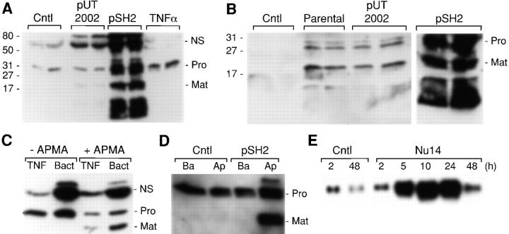
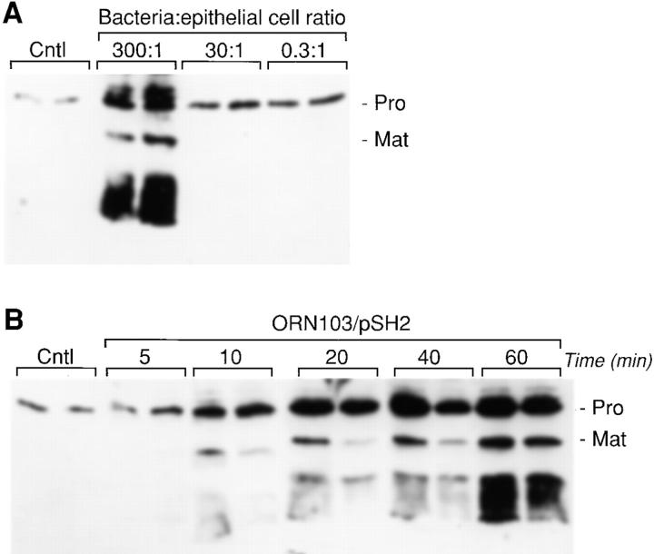
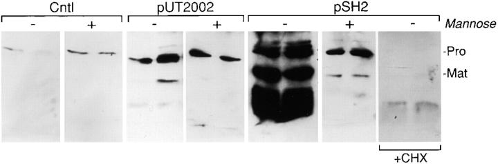
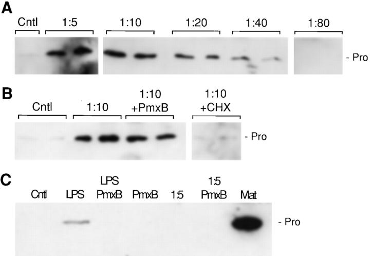
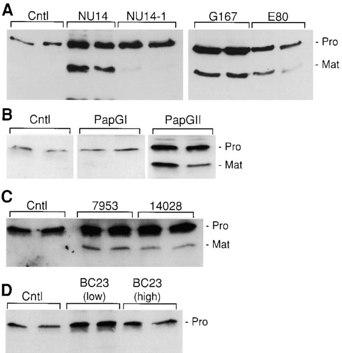
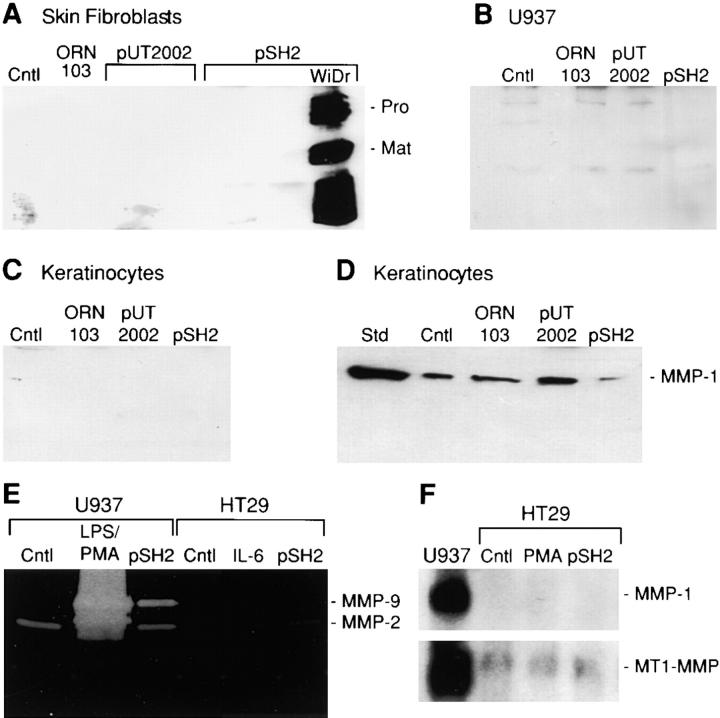
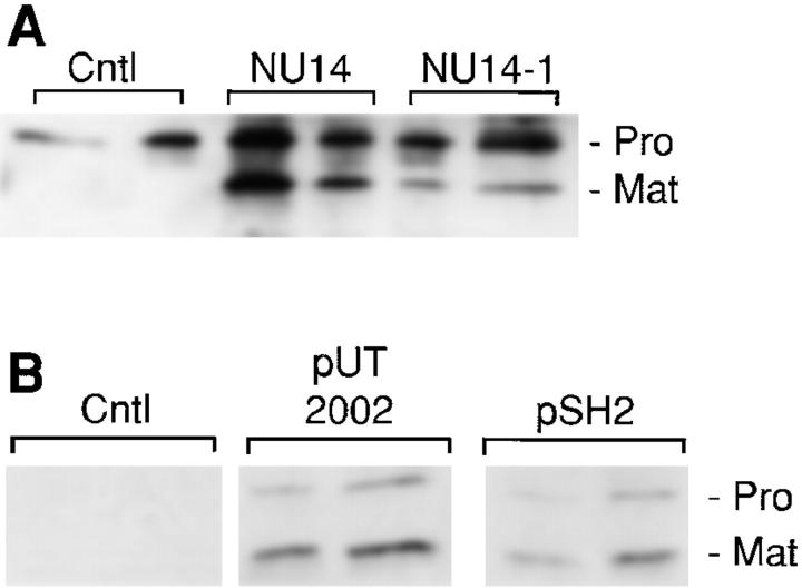
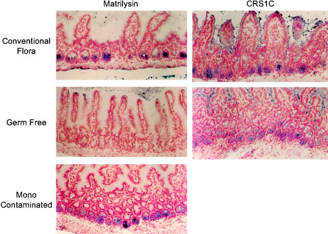
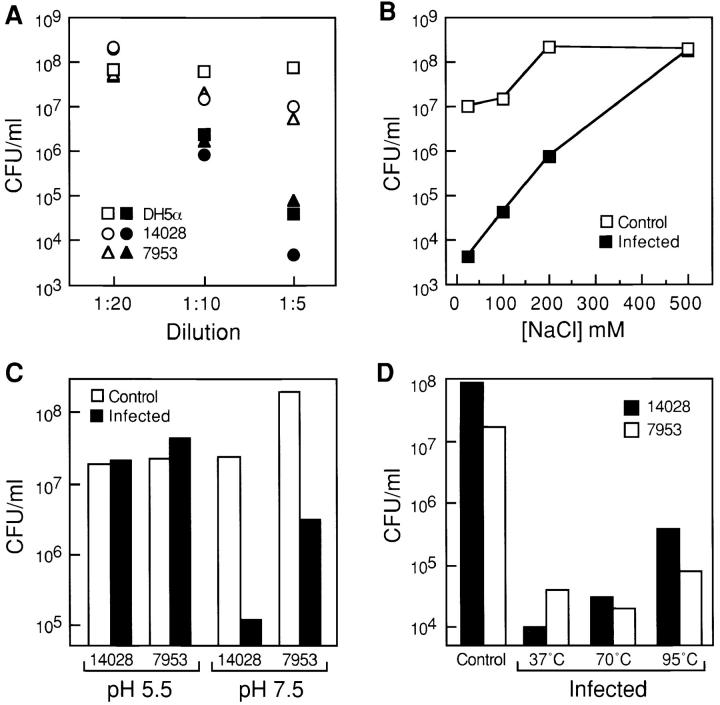
Matrilysin, a matrix metalloproteinase, is expressed and secreted lumenally by intact mucosal and glandular epithelia throughout the body, suggesting that its regulation and function are shared among tissues. Because matrilysin is produced in Paneth cells of the murine small intestine, where it participates in innate host defense by activation of prodefensins, we speculated that its expression would be influenced by bacterial exposure. Indeed, acute infection (10-90 min) of human colon, bladder, and lung carcinoma cells, primary human tracheal epithelial cells, and human tracheal explants with type 1-piliated Escherichia coli mediated a marked (25-50-fold) and sustained (>24 h) induction of matrilysin production. In addition, bacterial infection resulted in activation of the zymogen form of the enzyme, which was selectively released at the apical surface. Induction of matrilysin was mediated by a soluble, non-LPS bacterial factor and correlated with the release of defensin-like bacteriocidal activity. Bacteria did not induce matrilysin in other cell types, and expression of other metalloproteinases by epithelial cells was not affected by bacteria. Matrilysin was not detected in germ-free mice, but the enzyme was induced after colonization with Bacteroides thetaiotaomicron. These findings indicate that bacterial exposure is a potent and physiologically relevant signal regulating matrilysin expression in epithelial cells.


Figures

Figure 1
Figure 1
FimH-dependent induction and activation of matrilysin in epithelial cells. (A) WiDr human colon carcinoma cells were infected for 90 min with E. coli strains ORN103/pUT2002 (type 1+/fimH ) and ORN103/pSH2 (type 1+/fimH +) at a ratio of 300:1 bacteria/epithelial cells. Cultures were washed extensively and re-fed with medium containing 50 μg/ml gentamicin. Some cultures were treated for 48 h with 50 ng/ml of human recombinant TNFα. Conditioned media were collected 48 h after infection and analyzed by Western blotting for matrilysin protein. the 28-kD proform of matrilysin (Pro), the 19-kD activated form of matrilysin (Mat), and autolytic fragments (broad migrating faster below activated matrilysin) were seen in pSH2-infected cells. A nonspecific (NS) band is seen at ∼70 kD. Numbers on the left side of the autoradiogram indicate the migration of molecular mass standards. Samples from two separate cultures were analyzed for each group. (B) J82 human bladder carcinoma cells were infected for 90 min with the parental E. coli strain ORN103 and the recombinant strains ORN103/pUT2002 and ORN103/pSH2. Conditioned media were collected 48 h later, and the expression of matrilysin was analyzed by Western blotting. (C) Aliquots of conditioned media from TNFα-stimulated (TNF) or ORN103/pSH2-infected (Bact) colon epithelial cells were incubated at 37°C for 1 h in the presence or absence of 1 mM APMA, and the products were analyzed by Western blotting. (D) WiDr colon cells were grown on Transwell inserts, and some cultures were infected with ORN103/pSH2. Conditioned media was collected after 48 h from the apical and basal compartments and analyzed by Western blotting. (E) HT29 colon cells were infected for 90 min with E. coli NU14 (300 bacteria per epithelial cell). Total RNA was isolated at the indicated times after the infection. Matrilysin mRNA was detected by Northern hybridization.
Figure 2
Figure 2
Dose dependence and time-course analysis of bacterially induced upregulation of matrilysin. (A) HT29 human colon epithelial cells were infected for 90 min with E. coli strain ORN103/pSH2 (type 1+/fimH +) at the indicated bacterial/epithelial cell ratio. (B) HT29 cells were infected with the recombinant strain ORN103/pSH2, at a ratio of 300 bacteria per epithelial cell, for different periods of time, from 5 to 60 min. conditioned medium samples were collected at 48 h after infection and analyzed by Western blotting. (Pro) 28-kD proform of matrilysin; and (Mat) 19-kD activated matrilysin.
Figure 3
Figure 3
Mannose inhibits FimH-mediated induction and activation of matrilysin. WiDr human colon epithelial cells were infected for 90 min with the recombinant strains ORN103/pUT2002 (type 1+/fimH ) and ORN103/pSH2 (type 1+/fimH +) at a ratio of 300 bacteria per epithelial cell, in the presence or absence of 100 mM mannose or 10 μg/ml cycloheximide (CHX). Conditioned media were collected after a 48-h incubation and analyzed by Western blotting. (Pro) 28-kD proform of matrilysin; and (Mat) 19-kD activated matrilysin.
Figure 4
Figure 4
Inductive activity in bacterial conditioned medium. (A) HT29 human colon cells were treated with different dilutions of bacterial broth from the strain AAEC185/pSH2 (type 1+/fimH +) for 48 h, and matrilysin was assayed by Western blotting. (Cntl) Unstimulated cells; and (Pro) 28-kD promatrilysin. (B) HT29 cell cultures were incubated in duplicate with a 1:10 dilution of bacterial broth from the strain AAEC185/pSH2, in the presence or absence of 103 U/ml polymyxin B (Pmx B) and 10 μg/ml cycloheximide (CHX). (C) U937 human monocytic cells were incubated in the presence of 1 μg/ml E. coli LPS, and a 1:5 dilution of bacterial broth, in the presence or absence of 103 U/ml polymyxin B. MMP-7 denotes an aliquot of conditioned media from HT29 cells stimulated with bacterial broth.
Figure 5
Figure 5
Induction of matrilysin by adherent clinical isolates of E. coli and other bacteria. (A) WiDr human colon epithelial cells were infected for 60 min at a ratio of 300 bacteria per epithelial cell. The bacteria used were the type 1–piliated strains NU14, the isogenic FimH-deficient strain NU14-1, and the isolates G167 and EC80. (B) HT29 cells were infected with E. coli expressing type P pili and either the papGI or papGII adhesin. (C) HT29 cells were infected with S. typhimurium strains 14028s and ms7953s. (D) HT29 cells were infected with B. pertussis BC23 at two different ratios of bacteria to epithelial cell: 50:1 (low) and 500:1 (high). Western blotting was done with media collected 48 h after infection.
Figure 6
Figure 6
Cell type specificity of bacterial induction of matrilysin and lack of induction of other MMPs in epithelial cells. Human skin fibroblasts (A), U937 human monocytic-like cells (B), and primary human keratinocytes (C) were infected with the parental strain ORN103 and the type 1–piliated strains ORN103/pUT2002 (type 1+/fimH ) and ORN103/pSH2 (type 1+/fimH +). Western blotting for matrilysin (A, B, and C; MMP-7) and collagenase-1 (MMP-1; D) was performed on conditioned media collected 48 h after infection. WiDr human colon epithelial cells infected with ORN103/pSH2 were included as positive controls (A). (Cntl) Uninfected cells; and (Std) purified human collagenase-1. (E) Gelatin zymography was performed on serum-free conditioned media samples from infected U937 and HT29 cells. Zones of substrate clearing correspond to the activities of gelatinase-A (MMP-2) and gelatinase-B (MMP-9). A strong induction of gelatinase-B was observed in U937 cells treated for 24 h with 1 μg/ml E. coli LPS and 80 nM PMA. No gelatinolytic activity was seen in gels incubated in the presence of 50 mM EDTA (data not shown). (F) Northern blot analysis for collagenase-1 (MMP-1) and MT1-MMP mRNAs was performed using total RNA isolated from HT29 cells at 6 h after infection. RNA from U937 cells stimulated with LPS and PMA was used as a positive control for the expression of these MMPs. Ethidium bromide staining verified equal loading among lanes before and after transfer.
Figure 7
Figure 7
Ex vivo infection of human tracheal explants and infection of human tracheal epithelial cells. (A) Pieces (1 cm3) of freshly isolated normal adult human trachea were infected with the E. coli clinical isolates NU14 (fimH +) and NU14-1 (fimH ) for 90 min, washed extensively to eliminate nonadherent bacteria, and incubated for 24 h in fresh media containing 50 μg/ml gentamicin. Released matrilysin was detected by Western analysis. (B) Human tracheal primary epithelial cells were infected with the type 1–piliated recombinant strains ORN103/pSH2 (fimH +) and ORN103/pUT2002 (fimH ) for 90 min, and allowed to condition fresh media for 48 h. Matrilysin secretion was assessed by immunoblotting.
Figure 8
Figure 8
Bacterial-mediated induction of matrilysin in mouse small intestine. Immunostaining for mouse matrilysin and proCRS1C was done on frozen sections of the small intestine prepared from conventionally housed, germ-free, and ex-germ-free NMRI mice. Positive staining for matrilysin (blue) was seen in the Paneth cells located at the base of the intestinal crypts in mice with a normal intestinal microflora but was not detected in the small intestine of germ-free mice. Paneth cell expression of matrilysin was induced within 10 d after contamination with B. thetaiotaomicron. No changes in the staining pattern or intensity of procryptdin-related sequences (proCRS1C), a marker of Paneth cell differentiation, were seen in any samples studied.
Figure 9
Figure 9
Characterization of bacteriocidal activity in the epithelial cell conditioned media. (A) E. coli DH5α and S. typhimurium strains 14028s and ms7953s were incubated for 3 h at 37°C in the presence of different dilutions of conditioned medium from HT29 human colon epithelial cells, which were previously infected with the ORN103/pSH2 (type 1+/fimH +) strain (solid symbols), and from uninfected HT29 cells (open symbols). The number of colony forming units (CFU) was determined by plating serial dilutions of the cultures. The input number of bacteria was 2 × 10⁷ (DH5α), 2 × 10⁶ (14028s), and 106 (ms7953s) CFU/ml, respectively. (B) DH5α bacteria (2 × 10⁶ CFU/ml) were incubated for 3 h at 37°C in a 1:5 dilution of conditioned medium from bacterially infected or control HT29 cells containing increasing concentrations of NaCl. Serial dilutions were plated, and the number of colonies was determined after an overnight incubation at 37°C. (C) S. typhimurium 14028s (107 CFU/ml) and ms7953s (2.6 × 10⁷ CFU/ml) cells were incubated in the presence of a 1:5 dilution of conditioned medium from bacterially infected HT29 cells at pH 7.5 (50 mM phosphate buffer) or pH 5.5 (50 mM acetate buffer). (D) S. typhimurium 14028s and ms7953s cells were incubated with a 1:5 dilution of HT29 conditioned medium that was preincubated at 70°C for 30 min or at 95°C for 5 min.

Similar articles

Cited by

References

    1. Bals R., Goldman M.J., Wilson J.M. Mouse beta-defensin 1 is a salt-sensitive antimicrobial peptide present in epithelia of the lung and urogenital tract Infect. Immun. 66 1998. 1225 1232a - PMC - PubMed
    1. Bals R., Wang X., Wu Z., Freeman T., Bafna V., Zasloff M., Wilson J.M. Human β-defensin 2 is a salt-sensitive peptide antibiotic expressed in human lung J. Clin. Invest. 102 1998. 874 880b - PMC - PubMed
    1. Bals R., Wang X., Zasloff M., Wilson J.M. The peptide antibiotic LL-37/hCAP-18 is expressed in epithelia of the human lung where it has broad antimicrobial activity at the airway surface Proc. Natl. Acad. Sci. USA 95 1998. 9541 9546c - PMC - PubMed
    1. Birkedal-Hansen H. Role of matrix metalloproteinases in human periodontal diseases. J. Periodontol. 1993;64:474–484. - PubMed
    1. Birkedal-Hansen H., Moore W.G.I., Bodden M.K., Windsor L.J., Birkedal-Hansen B., DeCarlo A., Engler J.A. Matrix metalloproteinasesa review. Crit. Rev. Oral Biol. Med. 1993;4:197–250. - PubMed

Publication types

MeSH terms