. 2000 Mar 28;97(7):3094-9.
doi: 10.1073/pnas.97.7.3094.

Accuracy of thymine-thymine dimer bypass by Saccharomyces cerevisiae DNA polymerase eta

Affiliations

Accuracy of thymine-thymine dimer bypass by Saccharomyces cerevisiae DNA polymerase eta

M T Washington et al. Proc Natl Acad Sci U S A. .

Abstract

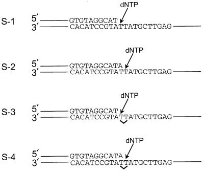
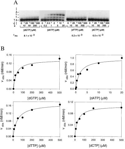
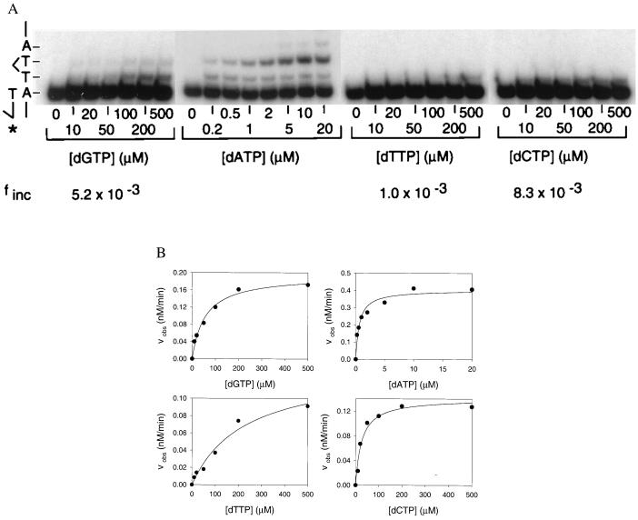
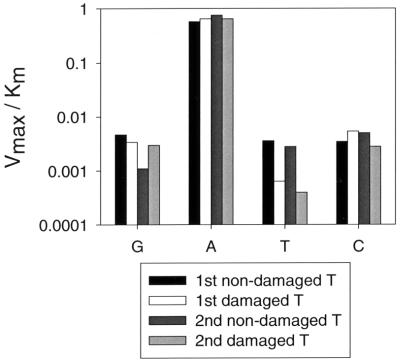
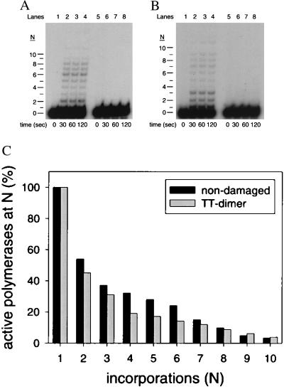
The Saccharomyces cerevisiae RAD30 gene functions in error-free replication of UV-damaged DNA. RAD30 encodes a DNA polymerase, Pol eta, which inserts two adenines opposite the two thymines of a cis-syn thymine-thymine (T-T) dimer. Here we use steady-state kinetics to determine the accuracy of DNA synthesis opposite the T-T dimer. Surprisingly, the accuracy of DNA synthesis opposite the damaged DNA is nearly indistinguishable from that opposite nondamaged DNA, with frequencies of misincorporation of about 10(-2) to 10(-3). These studies support the hypothesis that unlike most DNA polymerases, Pol eta is able to tolerate distortions in DNA resulting from damage, which then enables the polymerase to utilize the intrinsic base pairing ability of the T-T dimer.


Figures

Figure 1
Figure 1
DNA substrates used to assay Pol η fidelity. The nondamaged and damaged DNA substrates are shown. The position of the cis-syn T–T dimer in substrates S-3 and S-4 is indicated by a ∨. The first T and second T of the cis-syn T–T dimer or the identical nondamaged sequence refer to the first and second T residues encountered by the DNA polymerase by using that strand as a template.
Figure 2
Figure 2
Fidelity of Pol η opposite the first nondamaged template T. (A) Deoxynucleotide incorporation across from the first of two adjacent, nondamaged template T bases (substrate S-1; Fig. 1). Pol η (1 nM) was incubated for 5 min at 25°C with the primer-template DNA (10 nM) and with increasing concentrations of the incorrect deoxynucleotide (dGTP, dTTP, and dCTP; 0 to 500 μM) or the correct deoxynucleotide (dATP; 0 to 20 μM). The reactions were stopped and examined by denaturing PAGE. A portion of the template sequence is shown on the left. The asterisk indicates the 32P-labeled 5′ end of the primer. (B) Quantitation of deoxynucleotide incorporation reactions. The observed rate of deoxynucleotide incorporation is plotted as a function of concentration for each of the deoxynucleotides. The data were fit by using Eq. 1, and the Vmax and Km parameters obtained from the fit are listed in Table 1.
Figure 3
Figure 3
Fidelity of Pol η opposite the first damaged template T of the cis-syn T–T dimer. (A) Deoxynucleotide incorporation across from the first template T of the T–T dimer (substrate S-3; Fig. 1). Pol η (1 nM) was incubated for 5 min at 25°C with the primer-template DNA (10 nM) and with increasing concentrations of the incorrect deoxynucleotide (dGTP, dTTP, and dCTP; 0–500 μM) or the correct deoxynucleotide (dATP; 0–20 μM). The reactions were stopped and examined by denaturing PAGE. A portion of the template sequence is shown on the left. The asterisk indicates the 32P-labeled 5′ end of the primer. formula image T–T dimer. (B) Quantitation of the deoxynucleotide incorporation reactions. The observed rate of deoxynucleotide incorporation is plotted as a function of concentration for each of the deoxynucleotides. The data were fit by using Eq. 1, and the Vmax and Km parameters obtained from the fit are listed in Table 1.
Figure 4
Figure 4
Comparison of deoxynucleotide incorporation opposite the nondamaged and T–T dimer template residues. The Vmax/Km parameters (y axis) listed in Table 1 are shown for G, A, T, and C incorporation (x axis) opposite each of the two nondamaged and two damaged template T residues.
Figure 5
Figure 5
Processivity of Pol η on nondamaged and damaged substrates. (A) Processive DNA synthesis by Pol η on an undamaged DNA template (substrate S-1; Fig. 1) resulting from a single binding event. In lanes 1–4, Pol η (20 nM) was preincubated with the undamaged DNA substrate (10 nM) for 15 min at 25°C, and reactions were initiated by the addition of all four dNTPs (200 μM each), MgCl2 (5 mM), and the sonicated herring sperm DNA trap (1 mg/ml). Reactions were stopped after the indicated times, and the samples were examined by denaturing PAGE. The unextended primer (n = 0) and the extended primers (n = 1–10) are indicated. In lanes 5–8, Pol η was preincubated with the DNA substrate and with the DNA trap, and the reaction was initiated by the addition of dNTPs and MgCl2. (B) Processive DNA synthesis by Pol η on the damaged DNA template (substrate S-3; Fig. 1) resulting from a single DNA-binding event. Reactions were performed as described for the undamaged DNA template. (C) Percentage of active Pol η molecules at each position along the template. The black bars represent the active polymerases on the nondamaged DNA template and the gray bars represent the active polymerases on the damaged template.

Comment in

References

    1. Sancar A. Annu Rev Biochem. 1996;65:43–81. - PubMed
    1. Tang M, Bruck I, Eritja R, Turner J, Frank E G, Woodgate R, O'Donnell M, Goodman M F. Proc Natl Acad Sci USA. 1998;95:9755–9760. - PMC - PubMed
    1. Reuven N B, Tomer G, Livneh Z. Mol Cell. 1998;2:191–199. - PubMed
    1. Nelson J R, Lawrence C W, Hinkle D C. Science. 1996;272:1646–1649. - PubMed
    1. Johnson R E, Prakash S, Prakash L. Science. 1999;283:1001–1004. - PubMed

Publication types