. 2000 Mar 28;97(7):3649-54.
doi: 10.1073/pnas.97.7.3649.

A retinitis pigmentosa GTPase regulator (RPGR)-deficient mouse model for X-linked retinitis pigmentosa (RP3)

Affiliations

A retinitis pigmentosa GTPase regulator (RPGR)-deficient mouse model for X-linked retinitis pigmentosa (RP3)

D H Hong et al. Proc Natl Acad Sci U S A. .

Abstract

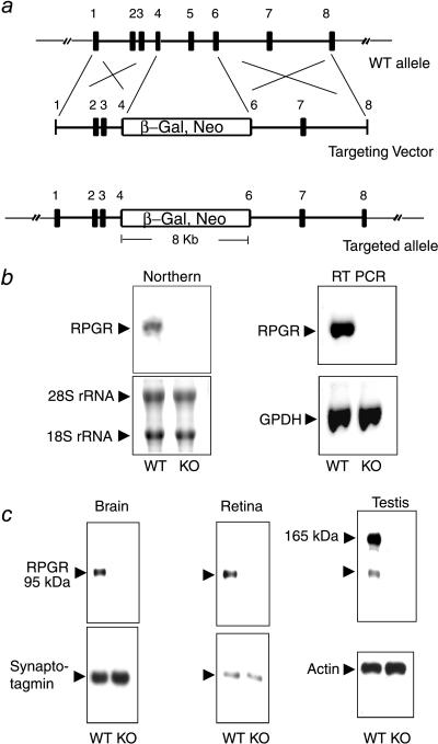
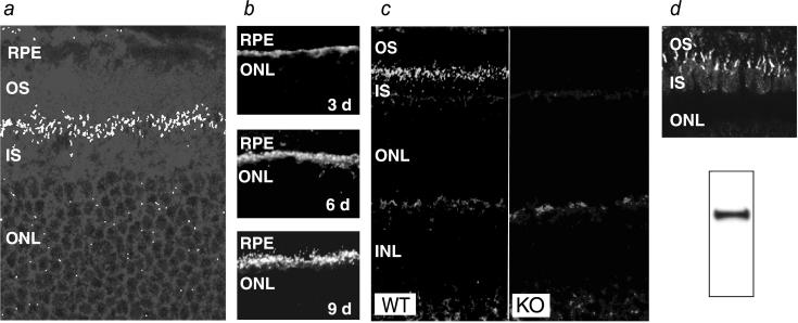
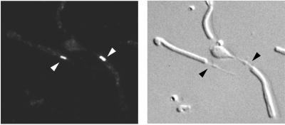
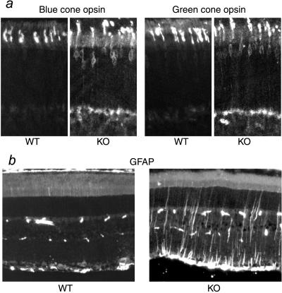
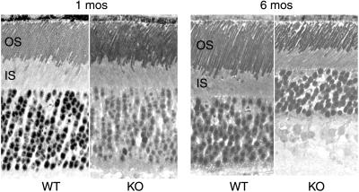
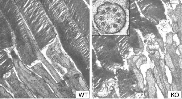
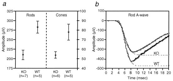
The X-linked RP3 locus codes for retinitis pigmentosa GTPase regulator (RPGR), a protein of unknown function with sequence homology to the guanine nucleotide exchange factor for Ran GTPase. We created an RPGR-deficient murine model by gene knockout. In the mutant mice, cone photoreceptors exhibit ectopic localization of cone opsins in the cell body and synapses and rod photoreceptors have a reduced level of rhodopsin. Subsequently, both cone and rod photoreceptors degenerate. RPGR was found normally localized to the connecting cilia of rod and cone photoreceptors. These data point to a role for RPGR in maintaining the polarized protein distribution across the connecting cilium by facilitating directional transport or restricting redistribution. The function of RPGR is essential for the long-term maintenance of photoreceptor viability.


Figures

Figure 1
Figure 1
Targeted disruption of the RPGR gene. (a) The targeting construct. Partial structure of the WT allele is shown (exons 1–8; filled boxes). The targeting vector contains a selectable marker (β-Gal, Neo) that replaces part of exon 4 through part of exon 6. (b) Absence of the normal RPGR transcript in the KO mice. (Left) Northern blot analysis with a full-length RPGR cDNA probe could detect a 3-kb transcript in brain RNA from WT but not from KO mice. Fluorescent dye-stained rRNA bands are shown as a loading control. (Right) Reverse transcription–PCR analysis confirms the absence of the normal RPGR transcript in the KO mice. PCR amplification of GPDH from the same cDNA templates is shown as a control. (c) RPGR protein expression was ablated in the KO mice. Total proteins from brain, retina, or testis were probed with anti-RPGR antibodies on immunoblots. The same blots were reprobed with either synaptotagmin (abundant in neural tissues) or actin antibodies as loading controls.
Figure 2
Figure 2
Localization of RPGR in the connecting cilium by immunofluorescence. (a) Confocal image of a normal retina probed with the RPGR antibodies. A band of punctate staining is seen between the inner and outer segments, indicative of connecting cilia. (×500.) (b) Developmental time course of RPGR staining in the mouse retina. Staining first appears around postnatal day 3 as a band at the apex of the photoreceptor layer and increases in intensity and definition through day 9. (c) The connecting cilium staining for RPGR is absent in KO mice. (×250.) (d) RPGR is also localized in the connecting cilia of cone photoreceptors. Shown is RPGR immunostaining of the cone-dominant ground squirrel retina (Upper). Western blotting of ground squirrel tissue homogenates confirms that the RPGR antibodies made against mouse sequence cross-reacts with squirrel RPGR (Lower). RPE, retinal pigment epithelium; OS, outer segment; IS, inner segment; ONL, outer (photoreceptor) nuclear layer.
Figure 3
Figure 3
Immunostaining of dissociated photoreceptors provides higher spatial resolution. Shown are epifluorescence (Left) and Nomarski (Right) images of the same field of view. Arrows point to connecting cilia of rod photoreceptors. (×500.)
Figure 4
Figure 4
Histopathology in KO retinas revealed by immunocytochemistry. (a) Mislocalization of blue and green cone opsins in the KO retinas. (b) GFAP is up-regulated in the KO retinas. (Left) GFAP immunoreactivity is normally found only in the astrocytes in the ganglion cell layer of the retina (near the bottom of figure). The sporadic speckles in the middle of the figure are blood vessel profiles recognized by the anti-mouse secondary antibody. (Right) Muller cell processes are prominently stained for GFAP in a 2-month-old KO retina.
Figure 5
Figure 5
Light micrographs of retinal sections from WT and KO littermates at 1 and 6 months of age. Note the reduced outer segment length and reduced thickness of the outer nuclear layer in the KO retina at 6 months of age. (×300.)
Figure 6
Figure 6
Electron micrographs of photoreceptors from WT (Left) and KO (Right) mice. Both have well packed disks in the outer segments, but the newly added disk membranes at the base of the outer segments appear disorganized in the KO photoreceptors. (×16,000.) (Inset) Cross section through a connecting cilium showing the normal organization of 9 + 0 microtubule arrangement in the mutant photoreceptor. (×80,000.)
Figure 7
Figure 7
Rod and cone ERGs from WT and KO mice at 6.5 months of age. (a) Means and SEM of rod ERG a-wave amplitudes (Left) and cone ERG b-wave amplitudes (Right). (b) Representative dark-adapted ERG a-waves in response to a bright flash of white light (28 foot Lambert-seconds) from WT and KO mice. Dashed lines are fitted functions.

References

    1. Berson E L, Rosner B, Simonoff E. Am J Ophthalmol. 1980;89:763–775. - PubMed
    1. Beron E L, Gouras P, Gunkel R D, Myrianthopoulos N C. Arch Ophthalmol. 1969;81:215–225. - PubMed
    1. Jacobson S G, Buraczynska M, Milam A H, Chen C, Jarvalainen M, Fujita R, Wu W, Huang Y, Cideciyan A V, Swaroop A. Invest Ophthalmol Visual Sci. 1997;38:1983–1997. - PubMed
    1. Weleber R G, Butler N S, Murphey W H, Sheffield V C, Stone E M. Arch Ophthalmol. 1997;115:1429–1435. - PubMed
    1. Buraczynska M, Wu W, Fujita R, Buraczynska K, Phelps E, Andreasson S, Bennett J, Birch D G, Fishman G A, Hoffman D R, et al. Am J Hum Genet. 1997;61:1287–1292. - PMC - PubMed

Publication types