. 2000 Mar 28;97(7):3759-64.
doi: 10.1073/pnas.97.7.3759.

The integral membrane S-locus receptor kinase of Brassica has serine/threonine kinase activity in a membranous environment and spontaneously forms oligomers in planta

Affiliations

The integral membrane S-locus receptor kinase of Brassica has serine/threonine kinase activity in a membranous environment and spontaneously forms oligomers in planta

J L Giranton et al. Proc Natl Acad Sci U S A. .

Abstract

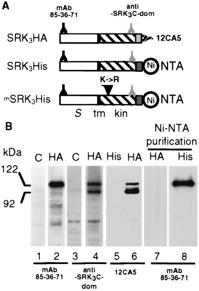
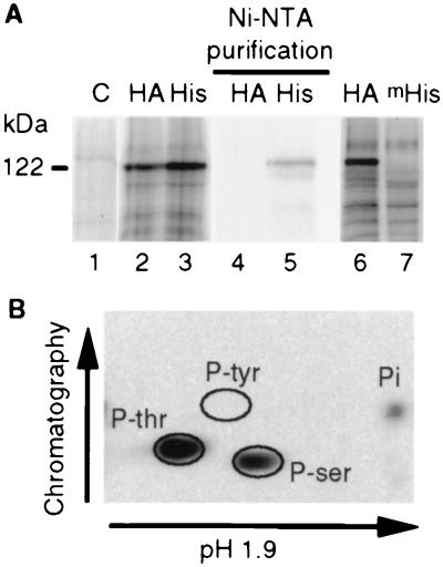
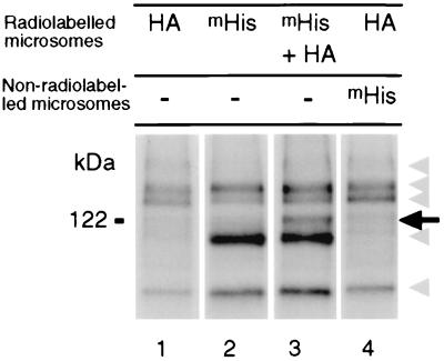
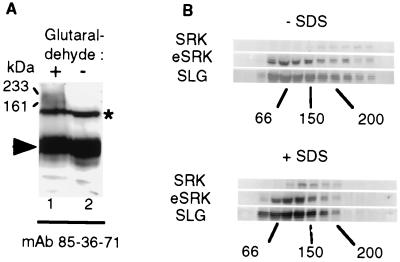
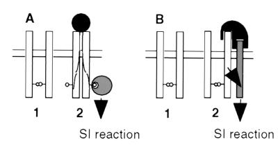
To gain further insight into the mode of action of S-locus receptor kinase (SRK), a receptor-like kinase involved in the self-incompatibility response in Brassica, different recombinant SRK proteins have been expressed in a membranous environment using the insect cell/baculovirus system. Recombinant SRK proteins exhibited properties close to those of the endogenous stigmatic SRK protein and were found to autophosphorylate on serine and threonine residues in insect cell microsomes. Autophosphorylation was constitutive because it did not require the presence of pollen or stigma extracts in the phosphorylation buffer. Phosphorylation was shown to occur in trans, suggesting the existence of constitutive homooligomers of membrane-anchored recombinant SRK. To investigate the physiological relevance of these results, we have examined the oligomeric status of SRK in planta in cross-linking experiments and by velocity sedimentation on sucrose gradients. Our data strongly suggest that SRK is associated both with other SRK molecules and other stigma proteins in nonpollinated flowers. These findings may have important implications for our understanding of self-pollen signaling.


Figures

Figure 1
Figure 1
Expression of recombinant SRK3 proteins in insect cells. (A) Schematic representation of the three recombinant SRK proteins, SRK3HA, SRK3His, and mSRK3His. The position of the different epitopes and tags are shown. The N and C termini of the recombinant proteins are to the left and right, respectively. The white rectangle represents the S-domain (S), the black vertical bar the membrane-spanning (tm) domain, the hatched rectangle indicates the cytoplasmic domain (kin) and the light and dark gray stippled rectangles indicate the HA epitope and hexahistidine tag, respectively. Epitopes recognized by different antibodies and the binding site for Ni-NTA are indicated. The substitution of Lys-553 with an arginine (K->R) in the mSRK3His construct is indicated by a vertical arrowhead. (B) Immunoblotting of SRK recombinant proteins. Proteins extracted from Sf21 cells infected by the parental baculovirus (C) (lanes 1 and 3) or from Sf21 cells infected with baculovirus driving the expression of SRK3HA (HA) (lanes 2, 4, 6, and 7) or SRK3His (His) (lanes 5 and 8) were separated by SDS/PAGE and electroblotted. In some cases, electrophoresis was preceded by a purification with Ni-NTA agarose beads as indicated.
Figure 2
Figure 2
Recombinant SRK autophosphorylates on serine and threonine residues in a membranous environment. (A) Microsomes from uninfected Sf21 cells (C) or from Sf21 cells expressing SRK3HA (HA), SRK3His (His) or kinase defective mSRK3His (mHis) were radiolabeled with [γ-32P]ATP. In some cases, proteins (lanes 4 and 5) were purified on Ni-NTA agarose beads after radiolabeling. Proteins were separated by SDS/PAGE and detected by autoradiography. (B) Radiolabeled SRK3His was purified on Ni-NTA agarose beads and hydrolyzed. Free amino acids were separated in two dimensions by chromatography and electrophoresis as indicated, and radiolabeled amino acids were detected by autoradiography. Phosphoamino acids (P-ser, P-thr, and P-tyr for phosphoserine, phosphothreonine, and phosphotyrosine, respectively) were positioned by staining nonradiolabeled phosphoamino acids, added before separation, with nihydrin. Pi indicates inorganic phosphate.
Figure 3
Figure 3
Intermolecular phosphorylation of recombinant SRK proteins. Microsomes from Sf21 cells expressing either SRK3HA (HA) or kinase-defective mSRK3His (mHis) or from Sf21 cells coexpressing mSRK3His and SRK3HA (mHis + HA) were radiolabeled, and proteins carrying the hexahistidine tag were purified on Ni-NTA agarose beads. For the sample shown in lane 4, extracts of unlabeled microsomes from cells expressing mSRK3His were added before purification on Ni-NTA agarose beads. Proteins eluted from Ni-NTA agarose beads were separated by SDS/PAGE and analyzed by autoradiography. Truncated recombinant SRK3 and contaminating polypeptides are indicated by gray arrowheads, and phosphorylated SRK is indicated by an arrow.
Figure 4
Figure 4
Identification of SRK oligomeric complexes in stigma extract. (A) Proteins were extracted from stigmas expressing the S3 haplotype in a Triton X-100-containing buffer and either treated (+) or not (−) with the cross-linking reagent glutaraldehyde. Proteins then were separated by SDS/PAGE and immunoblotted with mAb 85–36-71. SRK3-containing complexes of 161 and 233 kDa are indicated by arrows. Monomeric SRK3 and eSRK3 are indicated with an asterisk and an arrowhead, respectively. (B) Velocity sedimentation on sucrose gradients. Proteins were extracted in buffer containing octyl-glucoside, and stigma extracts were either supplemented with SDS at the concentration of 0.5% (mass/vol) (+SDS) or not (−SDS). The presence of SRK3 (SRK), eSRK3 (eSRK), and SLG3 (SLG) in the different fractions was examined by immunoblotting with mAb 85–36-71 and anti-SLG3 antibodies. The distribution of molecular mass markers (66, 150, and 200 kDa) is indicated at the bottom of each panel.
Figure 5
Figure 5
Two models for the molecular mechanism of signal transduction via SRK in the SI response. (A) This model proposes that SRK (open rectangle) spontaneously associates as a dimer in the plasmalemma of stigmatic papillar cells before pollination. Interaction with a self-pollen borne ligand (filled circle) induces a conformational change of SRK, which allows the recruitment of cytoplasmic targets (large hatched circles) that mediate the SI response. In the example shown here, the cytoplasmic targets recognize phosphorylated residues (small circles) on the SRK protein. (B) In the second model, SRK is also present as a constitutively formed dimer but the activation of SI response by pollen-ligand (in black) requires both binding to SRK in an allele-specific manner, and then binding to a second as-yet-unknown coreceptor (hatched rectangle), which does not need to exhibit any allelic specificity. Signal transduction then may involve interaction of the second receptor with cytosolic targets analogous to those described for serine/threonine receptor kinases in animals.

Similar articles

Cited by

References

    1. De Nettancourt D. Incompatibility in Angiosperms. New York: Springer; 1977.
    1. Nasrallah J B, Kao T-H, Goldberg M L, Nasrallah M E. Nature (London) 1985;318:617–618.
    1. Stein J C, Howlett B, Boyes D C, Nasrallah M E, Nasrallah J B. Proc Natl Acad Sci USA. 1991;88:8816–8820. - PMC - PubMed
    1. Kandasamy M K, Paolillo D J, Faraday C D, Nasrallah J B, Nasrallah M E. Dev Biol. 1989;134:462–472. - PubMed
    1. Delorme V, Giranton J L, Hatzfeld Y, Friry A, Heizmann P, Ariza M J, Dumas C, Gaude T, Cock J M. Plant J. 1995;7:429–440. - PubMed

Publication types

MeSH terms