. 2000 Mar 28;97(7):3608-13.
doi: 10.1073/pnas.97.7.3608.

Differential regulation of circadian pacemaker output by separate clock genes in Drosophila

Affiliations

Differential regulation of circadian pacemaker output by separate clock genes in Drosophila

J H Park et al. Proc Natl Acad Sci U S A. .

Abstract

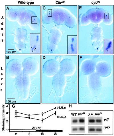
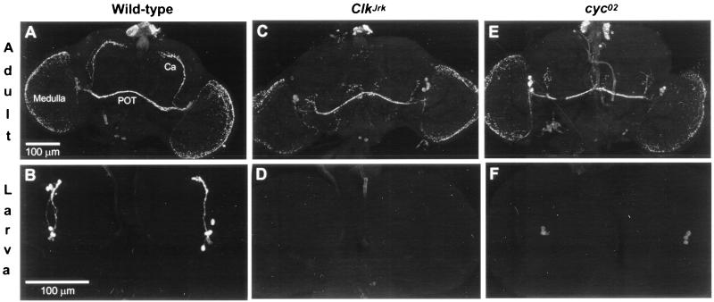
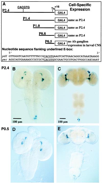
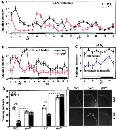
Regulation of the Drosophila pigment-dispersing factor (pdf) gene products was analyzed in wild-type and clock mutants. Mutations in the transcription factors CLOCK and CYCLE severely diminish pdf RNA and neuropeptide (PDF) levels in a single cluster of clock-gene-expressing brain cells, called small ventrolateral neurons (s-LN(v)s). This clock-gene regulation of specific cells does not operate through an E-box found within pdf regulatory sequences. PDF immunoreactivity exhibits daily cycling, but only within terminals of axons projecting from the s-LN(v)s. This posttranslational rhythm is eliminated by period or timeless null mutations, which do not affect PDF staining in cell bodies or pdf mRNA levels. Therefore, within these chronobiologically important neurons, separate elements of the central pacemaking machinery regulate pdf or its product in novel and different ways. Coupled with contemporary results showing a pdf-null mutant to be severely defective in its behavioral rhythmicity, the present results reveal PDF as an important circadian mediator whose expression and function are downstream of the clockworks.


Figures

Figure 1
Figure 1
Spatial and temporal expression of pdf RNA. Whole-mounted nervous systems were processed for pdf in situ hybridization. (A) Wild-type (Canton-S) adults (n = 5 dissected CNSs). Almost invariably, there were four signal-containing s-LNvs and four l-LNvs in each of the 10 brain hemispheres. The boxed area is a 4× magnification of the lateral-brain region; asterisked cells are the s-LNvs. (B) Wild-type larvae (n = 6); four signal-containing brain cells were invariably stained in each of the 12 brain hemispheres. (C) ClkJrk mutant adults (n = 7); there were no discernible s-LNvs in any of the 14 hemispheres. (D) ClkJrk larvae (n = 6); no pdf-RNA-containing cells were detected in the 12 hemispheres. (E) cyc0 mutant adults. The results depicted here for cyc02 (n = 7) were very similar those obtained from cyc01 (n = 8); in both allelic mutant types, there were subnormal numbers of s-LNvs labeled by the pdf probe; this is exemplified by the one asterisked cell in the 4×-magnified box for the lateral-brain region of this cyc02 adult (compare with the box in A); the reductions in s-LNv cell numbers in cyc0 adults were best appreciated after immunohistochemistry (see Fig. 2). (F) cyc0 larvae; for cyc02 (as exemplified in this image), 2.6 ± 0.3 lateral neurons (LNs) were labeled per hemisphere (n = 14), and these RNA signals were weak compared with wild-type; corresponding values for cyc01: 3.4 ± 0.2 (n = 8). Near the bottom of A–F is the abdominal ganglion, within which there is a small cluster of pdf cells. (G) Timecourse of pdf mRNA in separate clusters of peptide-containing neurons; in situ-hybridized specimens were blind-scored for staining levels, using 7–16 adult brains per time-point (the scoring scale ranged from 0 to 4, but only 3% of the individual values ranged as low as 1); ZTs are Zeitgeber times in which light comes on at noon during a 12-h light (open bar)–12-h dark (black bar) cycle. (H) pdf RNA levels in period and timeless mutants; the Northern-blot results were obtained from head extracts after sacrificing adults at ZT2–4; normalizing the per+ and tim+ levels (set = 1.00) to the rp49 loading control, the abundance in per01 was 1.07 and that for tim01 was 0.95. In situ hybridizations on y per01 w, y w;tim01, and y w control brains (dissected from adults at ZT2–5) led to the following staining scores (as on the ordinate of G): s-LNvs, in the genotypic order just given: 2.1 ± 0.2 (n = 16 brain hemispheres), 1.8 ± 0.3 (n = 10), 2.4 ± 0.2 (n = 8); l-LNvs: 3.0 ± 0.2, 2.7 ± 0.3, 3.6 ± 0.2; by ANOVA, the differences among s-LNv scores were not significant (P = 0.1); those among l-LNv scores were significant (P = 0.03), but only one pairwise comparison was marginally so (tim01 vs. control, P ≈ 0.05).
Figure 2
Figure 2
Effects of Clock and cycle mutations on PDF immunostaining in brain neurons. (A) Wild-type adults (n = 14); numbers of signal-containing cells per brain hemisphere: s-LNvs, 3.0 ± 0.2; l-LNvs, 3.6 ± 0.1 (n = 27 scorable hemispheres). Ca, calyx of the dorsal-brain mushroom body, in the vicinity of which are termini of neurites projecting from s-LNv cells; POT, posterior optic tract, one type of projection from l-LNv cells. (B) Wild-type larvae (n = 6); four PDF-containing LNs were invariably detected in the 12 brain hemispheres examined. (C) ClkJrk adults (n = 10); no s-LNvs were detected in the 20 brain hemispheres examined for mutant homozygotes, nor were there any such signals in 7 brains of flies heterozygous for ClkJrk and a deletion (Df) of the locus; counts of stained l-LNvs in ClkJrk/ClkJrk: 3.8 ± 0.2 (n = 17 scorable hemispheres). Arrow, abnormal projections. (D) ClkJrk larvae (n = 14); no LNs were detected in any of the 28 hemispheres examined, nor were any observed in the brains of 8 larvae heterozygous for ClkJrk and the Df. (E) cyc0 adults; this cyc02 specimen exemplifies the subnormalities and abnormalities of s-LNv cells exhibited by the 10 adults examined that were homozygous this allele—e.g., no PDF neurites extending toward the top of the brain. Arrowhead, abnormal projections. For the companion cyc01 mutant (n = 9 adult CNSs dissected and stained) cell counts: s-LNvs, 1.0 ± 0.2; l-LNvs, 3.4 ± 0.2 (n = 15 scorable hemispheres). (F) cyc0 larvae. Consistent with the in situ hybridizations (Fig. 1), the staining intensities in PDF cells were much weaker than in wild-type and variable within a given specimen; regardless of the number of LNs detected in either cyc01 or cyc02 larvae (n = 16 and 10, respectively), no stained axonal processes were detectable. Cell counts: cyc01, 3.1 ± 0.3 (n = 32 hemispheres); cyc02, 2.9 ± 0.3 (n = 20).
Figure 3
Figure 3
Analysis of the pdf promoter region. (A) E-box sequence ca. 1.4 kb upstream of the transcription start site (Tx) in the P2.4 construct. Half of the E-box (GTG) is present in P1.4 but eliminated in the other three constructs; note that there is no homology in the nucleotide sequences flanking per's and pdf's E-boxes (per upstream sequence: ref. 2). (B) 5-Bromo-4-chloro-3-indolyl β-d-galactoside (X-Gal) histochemistry for a P2.4 + UAS-lacZ larval CNS. (C) Adult CNS of the same transgenic type as in B. Note that in the imaginal specimen there are only LN signals, no dorsal signals (cf. Figs. 1 and 2). (D) X-Gal-mediated staining in a P0.5 + UAS-lacZ larval CNS. There is no reporter expression in the abdominal ganglion. (E) Adult CNS of the same transgenic type as in D.
Figure 4
Figure 4
Temporal analysis of PDF-like immunoreactivity in brain cells and their projections. (A) Immunohistochemical time-course of wild-type head sections and those of the perS and per01 mutants, in both LD-cycling and constant-dark (DD) conditions (n = 10–13 per time-point for each genotype). ZTs, as in Fig. 1; CT, circadian times during DD (gray bar, subjective day, corresponding to actual daytime in the preceding LD condition; black bar, subjective night). The apparent oscillations were formally analyzed for the DD portions of the records, resulting in 24.2-h and 19.9-h free-running period estimates for wild-type and perS, respectively. In per01 specimens, examined at eight equally spaced times in LD, the nerve-terminal signals were chronically low: at five time-points, the staining levels were 0; at the other three, the average scores ranged from 0.1 to 0.2. (B) s-LNv cell-body staining scores for the same specimens as in A. (C) l-LNv-cell staining for cell bodies and centrifugally projecting fibers; the specimens scored are a subset of those that led to the scoring for A and B (here, only the flies that were in the LD cycle). (D) Immunohistochemical comparison of wild type (WT) to two clockless mutants. The nerve-terminal signals were examined in wild type, per01, and tim01 at the peak and trough times (cf. A) in an LD cycle; these brains were whole-mounted, and the relevant dorsally located anti-PDH-mediated signals were quantified as described in the text; y w is a tim+ control for the arrhythmic loss-of-function timeless mutant (which was in a y w genetic background); based on the numerical read-outs from analysis of the nerve-terminal staining intensities, certain of the average signals were determined to be significantly different (P < 0.05), as indicated by ∗∗; the numbers of brain hemispheres examined are shown in parentheses; error bars, SEM. (E) Representative dorsal-brain images of s-LNv nerve terminals (cf. Fig. 2A), stained after sacrificing flies of three genotypes in the early morning (ZT1) or the early night (ZT13).

References

    1. Dunlap J C. Cell. 1999;96:271–290. - PubMed
    1. Hao H, Allen D, Hardin P E. Mol Cell Biol. 1997;17:3687–3693. - PMC - PubMed
    1. Darlington T K, Wager-Smith K, Ceriani M F, Staknis D, Gekakis N, Steeves T D L, Weitz C J, Takahashi J S, Kay S A. Science. 1998;280:1599–1603. - PubMed
    1. Lee C, Bae K, Edery I. Mol Cell Biol. 1999;19:5316–5325. - PMC - PubMed
    1. Jin X, Shearman L P, Weaver D R, Zylka M J, de Vries G J, Reppert S M. Cell. 1999;96:57–68. - PubMed

Publication types

LinkOut - more resources