. 2000 Mar 28;97(7):3376-81.
doi: 10.1073/pnas.97.7.3376.

The phytopathogenic bacteria Erwinia carotovora infects Drosophila and activates an immune response

Affiliations

The phytopathogenic bacteria Erwinia carotovora infects Drosophila and activates an immune response

A Basset et al. Proc Natl Acad Sci U S A. .

Abstract

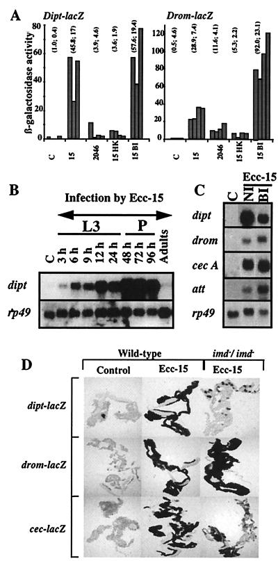
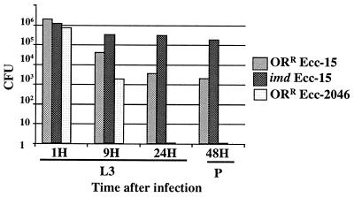
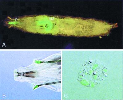
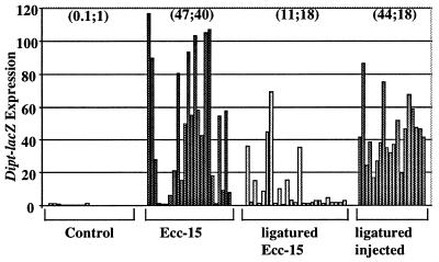
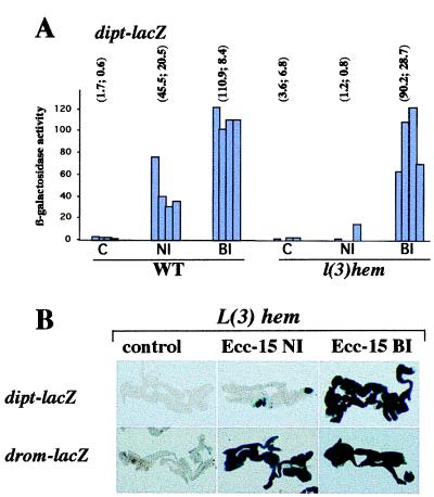
Although Drosophila possesses potent immune responses, little is known about the microbial pathogens that infect Drosophila. We have identified members of the bacterial genus Erwinia that induce the systemic expression of genes encoding antimicrobial peptides in Drosophila larvae after ingestion. These Erwinia strains are phytopathogens and use flies as vectors; our data suggest that these strains have also evolved mechanisms for exploiting their insect vectors as hosts. Erwinia infections induce an antimicrobial response in Drosophila larvae with a preferential expression of antibacterial versus antifungal peptide-encoding genes. Antibacterial peptide gene expression after Erwinia infection is reduced in two Drosophila mutants that have reduced numbers of hemocytes, suggesting that blood cells play a role in regulating Drosophila antimicrobial responses and also illustrating that this Drosophila-Erwinia interaction provides a powerful model for dissecting host-pathogen relationships.


Figures

Figure 1
Figure 1
Induction of antimicrobial peptide genes after Ecc-15 infection. (A) Viable Ecc-15 is required to induce a larval immune response. Quantitative measurements of β-galactosidase activity in larvae carrying either the dipt-lacZ (Left) or the drom-lacZ (Right) reporter genes were performed on five larvae collected 24 h after various treatments. Each bar represents an independent measurement; mean and SD are indicated above. C, control larvae incubated in banana alone; 15 and 2046, larvae naturally infected with Ecc-15 and Ecc-2046, respectively; 15-HK, heat-inactivated Ecc-15 (40 min at 70°C); 15-BI, larvae injected with Ecc-15 (bacterial injection). (B) Northern blot analysis shows that Ecc-15 infection induces sustained diptericin expression in larvae and pupae collected at different time intervals after natural infection by Ecc-15. L3, third instar larvae; P, pupae; C, control. (C) Northern blot analysis shows that Ecc-15 infection of larvae induces the expression of all of the antimicrobial genes. Total RNA was collected 24 h after infection, and the blot was hybridized successively with the following cDNA probes: diptericin (dipt), cecropin A1 (cec A), attacin (att), drosomycin (drom), and ribosomal protein 49 (rp49) as an internal control. C, control larvae incubated with only banana; NI, larvae incubated with Ecc-15 (natural infection); BI, third instar larvae injected with Ecc-15 (bacterial injection). (D) Ecc-15 induces a systemic immune response. Histochemical staining of β-galactosidase activity is shown in the fat body of wild-type (OrR) and imd homozygous larvae that were collected 24 h after infection and that carry the dipt-lacZ, cec-lacZ, or drom-lacZ reporter genes. Ecc-15 infection induces high expression of all three reporter genes in fat bodies from wild-type larvae. The absence of β-galactosidase activity in fat bodies isolated from infected imd larvae carrying the dipt-lacZ and cec-lacZ genes indicates that the induction of cec-lacZ and dipt-lacZ expression by Ecc-15 requires the imd gene product.
Figure 2
Figure 2
Ecc-15 persists in developing larvae and pupae. Bacterial persistence was measured in wild-type (OrR) and imd/imd mutant lines by plating appropriate dilutions of homogenates of five surface-sterilized larvae or pupae that were naturally infected with rifampicin-resistant strains of Ecc-15 (Ecc-15 rifR) and Ecc-2046 (Ecc-2046 rifR) and collected at different times after infection. Bacterial counts were obtained by plating the larval and pupal homogenates on LB medium containing rifampicin (100 μg/ml; ref. 14). The number of colony-forming units (cfu) per larva obtained at each time point after infection represents the mean of three independent measurements. L3, third instar larvae; P, pupae.
Figure 3
Figure 3
Fate of Ecc-15 bacteria in the larvae. The localization of Ecc-15 within larvae was determined by using a strain of Ecc-15 (Ecc-15-GFP) that expresses the GFP reporter gene. (A) In a larva naturally infected by Ecc-15-GFP, bacteria are clearly present in the pharynx and anterior midgut. (B) In a larva infected by Ecc-15-GFP, bacteria are present in the anterior spiracles. (C) Hemolymph collected from larvae infected for 16 h with Ecc-15 shows GFP-marked bacteria phagocytosed by hemocytes. P, pharynx; AM, anterior midgut.
Figure 4
Figure 4
Ecc-15 infects larvae through the digestive tract. dipt-lacZ larvae that were sealed at the mouth by ligaturing with a strand of human hair were naturally infected by Ecc-15 for 12 h. Each bar represents the level of β-galactosidase activity measured in one larva. Means and sd are indicated above. Ligatured larvae generally failed to express dipt-lacZ after exposure to Ecc-15, indicating that the digestive tract is the major route of infection by Ecc-15. Ligatured larvae still have an wild-type immune response after Ecc-15 injection, indicating that the ligature blocks only the route of infection and does not alter host defense response. Control, untreated larvae; Ecc-15, larvae infected by Ecc-15; ligatured Ecc-15, ligatured larvae infected by Ecc-15; ligatured injected, ligatured larvae collected 8 h after Ecc-15 injection.
Figure 5
Figure 5
diptericin induction after natural infection by Ecc-15 requires hemocytes. (A) Quantitative measurements of β-galactosidase activity performed on five larvae collected 24 h after natural infection or bacterial injection by Ecc-15. Experiments were performed as described (3) on larvae carrying the dipt-lacZ reporter gene in either wild-type (WT) or l(3)hem/l(3)hem mutant backgrounds. C, control; NI, larvae incubated with Ecc-15 (natural infection); BI, third instar larvae injected with Ecc-15 (bacterial injection). Each bar represents an independent measurement. Means and sd are indicated above. (B) Histochemical staining of β-galactosidase activity is shown in the fat body of l(3)hem homozygous larvae carrying either the dipt-lacZ or drom-lacZ reporter genes. In contrast to injection, natural Ecc-15 infection failed to induce a strong expression of the dipt-lacZ reporter gene in the l(3)hem fat body. Larval fat bodies were isolated 24 h after infection and stained (5-bromo-4-chloro-3-indolyl β-d-galactoside) as described (11). C, unchallenged control larvae; Ecc-15 NI, natural infection by Ecc-15; Ecc-15 BI, bacterial injection of Ecc-15.

References

    1. Hoffmann J A, Reichhart J-M. Trends Cell Biol. 1997;7:309–316. - PubMed
    1. Hoffmann J A, Kafatos F C, Janeway C A, Ezekowitz R A B. Science. 1999;284:1313–1318. - PubMed
    1. Lemaitre B, Reichhart J M, Hoffmann J A. Proc Natl Acad Sci USA. 1997;94:14614–14619. - PMC - PubMed
    1. Agrios G N. Plant Pathology. 4th Ed. New York: Academic; 1997.
    1. Pérombelon M C M. Annu Rev Phytopathol. 1980;18:361–387.

Publication types

MeSH terms

LinkOut - more resources