

doi: 10.1073/pnas.070525997.
Influenza A and B viruses expressing altered NS1 proteins: A vaccine approach
Affiliations
- PMID: 10725408
- PMCID: PMC18238
- DOI: 10.1073/pnas.070525997
Item in Clipboard
Influenza A and B viruses expressing altered NS1 proteins: A vaccine approach
Proc Natl Acad Sci U S A.
.
Abstract
We propose a rational approach to the generation of live viral vaccines: alteration of virally encoded type I IFN antagonists to attenuate virulence while retaining immunogenicity. We have explored this concept by using the influenza virus. Previously we have shown that the NS1 protein of influenza A virus possesses anti-IFN activity. We now present evidence that influenza A and B viruses encoding altered viral NS1 proteins are highly attenuated in the mouse host, yet provide protection from challenge with wild-type viruses.
Figures
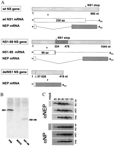
Wild-type A/PR8 influenza virus and derivative viruses containing alterations in the viral NS1 ORFs. (A) Schematic representation of the NS genes and gene transcripts for wild-type influenza A PR8 virus and transfectant NS1–99 and delNS1 viruses. Viral NS genes are indicated by light-gray boxes, with nucleotide length indicated in numbers below the gene segments. Stop codons for PR8 and NS1–99 NS1 ORFs are indicated by an asterisk above the viral genes. A 154-nt insertion in the NS1–99 gene is shown as a dark-gray box, and the 471-nt deletion of the delNS1 NS gene segment is represented by a vertical line with the deleted nucleotides listed below the viral gene. Viral NS1 ORFs are represented by white boxes with the amino acid length indicated within each box. Viral NEP mRNAs are also shown, with white boxes indicating the in-frame mRNA sequence shared between viral NS1 and NEP ORFs, and spotted boxes representing the unique ORFs of the viral NEP mRNA transcripts. Influenza A/PR8 and A/delNS1 viral NS genes have been described previously (4, 35) and are shown together with the A/NS1–99 NS gene for comparison. (B) Immunoprecipitation of NS1 protein from NS1–99 infected cells. MDCK cells were infected with PR8 or NS1–99 virus as described in Materials and Methods. 35S-met-cys-labeled NS1 protein was immunoprecipitated from infected cell extracts by using rabbit polyclonal antisera against the NS1 protein and separated on a 17.5% SDS gel. Proteins were visualized by autoradiography. (C) Western blot analysis of NEP expression. Vero cells were infected for the indicated time points at an MOI of 2 with either NS1–99, delNS1, or PR8 virus. Cell extracts were probed with an antibody against the viral NEP (Upper) or NP (Lower) as described in Materials and Methods.
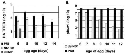
Growth characteristics of wild-type PR8, NS1–99, and delNS1 viruses in embryonated eggs. (A) One hundred pfu of virus was injected into the allantoic cavity of embryonated eggs of varying age and incubated for 48 h at 37°C. Allantoic fluid was harvested and titrated by HA assay as described in Materials and Methods. HA titers were not detected for delNS1 virus at days 8, 10, 12, or 14. NS1–99 infection did not give detectable HA titers at day 14. Graph represents the average reciprocal HA dilution of two to six eggs for each virus. (B) Embryonated eggs were infected with either PR8 or delNS1 virus as in A, and allantoic fluid was titrated on MDCK cells as described in Materials and Methods.
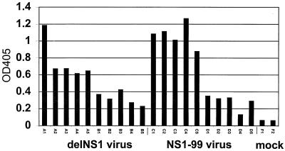
Influenza A-specific antibody titers of sera taken from mice immunized with delNS1 virus or NS1–99 virus. Serum samples were tested from sample groups A–D or from mock-immunized mice (group F) as indicated in Table 1. Samples were taken from mice 4 wk after immunization with primary virus and before challenge with wild-type PR8 virus. Samples were tested for their reactivity to sucrose purified influenza A/PR8. OD405 readings for serum diluted 1:1,000 are shown.

NP-peptide specific spleen cells from mice immunized with delNS1 virus or NS1–99 virus. Spleen cells from mice immunized with 1 × 10³ pfu of the indicated virus were harvested 10 days after immunization and assayed for influenza NP peptide-specific spleen cells, as described in Materials and Methods. Spots corresponding to IFN-γ-secreting cytotoxic T lymphocytes were counted at two different cell dilutions and averaged for each virus group. Results indicate the average number of spots of two different spleen cell dilutions, extrapolated to 1 × 10⁶ cells. Spleen cells were harvested from three mice for each group, with the exception of PR8-immunized mice. One of three mice infected with 1 × 10³ pfu of PR8 virus survived immunization and was killed for spleen cell analysis at day 10.
Wild-type B/Yamagata/1/73 influenza virus and derivative viruses containing alterations in the viral NS1 ORFs. Influenza B/Yamagata NS genes and mRNA transcripts are schematically represented as in Fig. 1. Viral NS gene segments are represented by light-gray boxes and viral NS1 ORFs by white boxes. A 13-nt deletion in the B/201 NS gene is indicated by a vertical bar. Seventeen amino acids resulting from a frameshift in the B/201 NS1 ORF are indicated by a hatched bar (8). Deletion of nucleotide number 310 in the B/234 NS gene segment results in a frameshift encoding one leucine and then a stop codon, represented by a vertical bar. The B/234 viral NS1 protein also contains a glu → val change at amino acid position 66 (not shown). B/Yamagata, B/201, and B/234 NS genes have been described previously, (6, 8) and are shown here together for comparison of the NS1 ORFs. Length of B/AWBY NS gene as predicted from the literature is indicated (6).
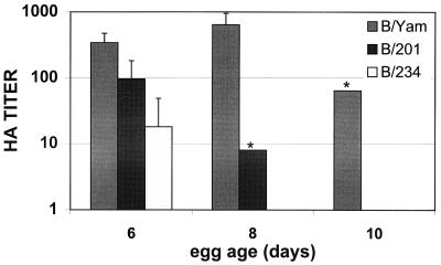
Growth characteristics of wild-type B/Yamagata, B/201, and B/234 viruses in embryonated eggs. One hundred pfu of influenza B viruses was injected into the allantoic cavity of embryonated eggs and incubated at 35°C for 72 h. Allantoic fluid was then harvested and subjected to HA analysis. Graph represents the average reciprocal HA dilution of two to six eggs for each virus. Asterisks indicate that two eggs were tested and gave the same HA titer. Influenza B/234 virus showed no HA titers in 8- and 10-day-old eggs.
Influenza B virus-specific antibody titers of sera taken from mice immunized with B/Yamagata, B/201, or B/234 virus. Serum samples were tested from mice immunized with 3 × 10⁵ pfu of the indicated virus or from mock immunized mice. Serum samples were taken from mice 4 wk after immunization and before challenge with wild-type B/Yamagata virus. Samples were tested for their reactivity against sucrose banded B/Yamagata virus. OD405 readings for sera diluted 1:1,000 are shown.
Similar articles
-
Development of a live-attenuated influenza B DeltaNS1 intranasal vaccine candidate.Vaccine. 2009 May 11;27(21):2851-7. doi: 10.1016/j.vaccine.2009.02.087. Epub 2009 Mar 11. Vaccine. 2009. PMID: 19366569
-
Growth and immunogenicity of influenza viruses cultivated in Vero or MDCK cells and in embryonated chicken eggs.Dev Biol Stand. 1999;98:39-51; discussion 73-4. Dev Biol Stand. 1999. PMID: 10494958
-
Immunogenicity and protection efficacy of replication-deficient influenza A viruses with altered NS1 genes.J Virol. 2004 Dec;78(23):13037-45. doi: 10.1128/JVI.78.23.13037-13045.2004. J Virol. 2004. PMID: 15542655 Free PMC article.
-
Identification of effective constituents of influenza vaccine by immunization with plasmid DNAs encoding viral proteins.Jpn J Infect Dis. 2000 Dec;53(6):219-28. Jpn J Infect Dis. 2000. PMID: 11227019 Review.
-
Learning from our foes: a novel vaccine concept for influenza virus.Arch Virol Suppl. 1999;15:131-8. doi: 10.1007/978-3-7091-6425-9_9. Arch Virol Suppl. 1999. PMID: 10470274 Review.
Cited by
-
The Dynamic Interface of Viruses with STATs.J Virol. 2020 Oct 27;94(22):e00856-20. doi: 10.1128/JVI.00856-20. Print 2020 Oct 27. J Virol. 2020. PMID: 32847860 Free PMC article. Review.
-
Evasion of influenza A viruses from innate and adaptive immune responses.Viruses. 2012 Sep;4(9):1438-76. doi: 10.3390/v4091438. Epub 2012 Sep 3. Viruses. 2012. PMID: 23170167 Free PMC article. Review.
-
Making better influenza virus vaccines?Emerg Infect Dis. 2006 Jan;12(1):61-5. doi: 10.3201/eid1201.051043. Emerg Infect Dis. 2006. PMID: 16494719 Free PMC article. Review.
-
The role of reverse genetics in the development of vaccines against respiratory viruses.Expert Opin Biol Ther. 2005 Mar;5(3):369-80. doi: 10.1517/14712598.5.3.369. Expert Opin Biol Ther. 2005. PMID: 15833074 Free PMC article. Review.
-
Extending the cytoplasmic tail of the influenza a virus M2 protein leads to reduced virus replication in vivo but not in vitro.J Virol. 2008 Jan;82(2):1059-63. doi: 10.1128/JVI.01499-07. Epub 2007 Nov 7. J Virol. 2008. PMID: 17989186 Free PMC article.
References
-
- Maassab H F, Herlocher M L, Bryant M L. In: Vaccines. Plotkin S A, Orenstein W A, editors. Philadelphia: Saunders; 1999. pp. 909–927.
-
- Gorse G J, Belshe R B. Scand J Infect Dis. 1991;23:7–17. - PubMed
-
- García-Sastre A, Egorov A, Matassov D, Brandt S, Levy D E, Durbin J E, Palese P, Muster T. Virology. 1998;252:324–330. - PubMed
-
- Luytjes W, Krystal M, Enami M, Pavin J D, Palese P. Cell. 1989;59:1107–1113. - PubMed
Publication types
MeSH terms
Substances
LinkOut - more resources
Full Text Sources
Other Literature Sources
Medical
