. 2000 Mar;105(6):741-8.
doi: 10.1172/JCI7997.

Gene transfer of the neuronal NO synthase isoform to cirrhotic rat liver ameliorates portal hypertension

Affiliations

Gene transfer of the neuronal NO synthase isoform to cirrhotic rat liver ameliorates portal hypertension

Q Yu et al. J Clin Invest. 2000 Mar.

Abstract

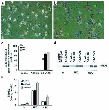
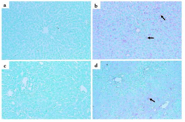
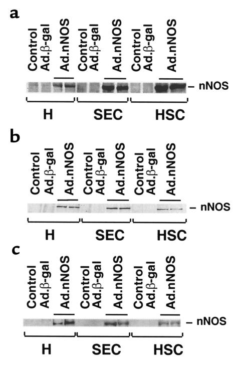
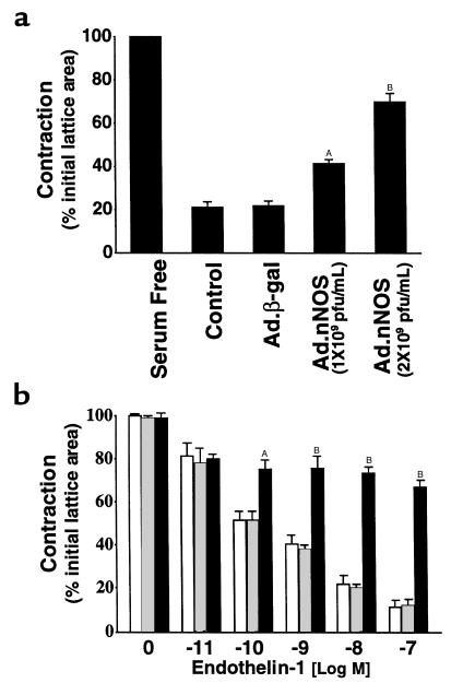
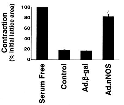
Reduced production of nitric oxide (NO) in the cirrhotic liver results from a defect in hepatic endothelial cell nitric oxide synthase (ecNOS) and appears to contribute to the high intrahepatic resistance and portal hypertension typical of cirrhosis. Therefore, we postulated that targeting a heterologous NOS isoform to sinusoidal endothelial cells or other perisinusoidal cells, such as hepatic stellate cells, would counter the defect in NO production and reduce resistance to blood flow. Recombinant adenovirus (Ad) carrying the neuronal NOS gene (nNOS) targeted liver sinusoidal endothelial cells, stellate cells, and hepatocytes more efficiently than the corresponding cells in cirrhotic livers, but transduction rates were substantial even in cirrhotic animals. Expression of nNOS in each liver cell type, whether from normal or injured liver, caused increased NO production and inhibited endothelin-1-induced contractility of perisinusoidal stellate cells. Finally, in 2 different in vivo models of cirrhosis and portal hypertension, transduction of livers with recombinant Ad.nNOS significantly reduced intrahepatic resistance and portal pressure. The data highlight the feasibility of gene transfer to diseased liver and hepatic cells and demonstrate the potential of a novel therapy for portal hypertension caused by cirrhosis.


Figures

Figure 1
Figure 1
Ad-mediated nNOS gene delivery and activity of nNOS. Parenchymal and nonparenchymal cells were prepared as in Methods. After isolation, cells were grown for 48–72 hours under standard conditions and then transduced with Ad.β-gal or Ad.nNOS as in Methods. In a and b, cells were transduced with Ad.β-gal or Ad.nNOS, respectively, and stained for NADPH diaphorase. In c, the transduction efficiency after exposure to Ad.nNOS was determined by cell counting as in Methods (Ad.β-gal is used as an additional control). In d, cell lysates (25 μg total protein) were immunoblotted for nNOS. In e, 24 hours after transduction, culture supernatants were collected and nitrite detected. AP < 0.01 compared with control or Ad.β-gal for each cell type (n = 3). H, hepatocyte; SEC, sinusoidal endothelial cells; HSC, hepatic stellate cells.
Figure 2
Figure 2
nNOS expression in vivo in normal and injured liver. Ad (1.5 × 10¹¹ pfu/kg via femoral vein) was administered to normal rats or those after liver injury as described in Methods. Liver tissue was fixed and probed for nNOS. In controls (a and c), immunoreactive nNOS is absent in normal and injured liver (BDL liver is shown; note the prominent bile duct proliferation) transduced with Ad.β-gal. In contrast, in both normal and injured livers transduced with nNOS (b and d), immunoreactive nNOS is identified in sinusoidal lining cells (arrows) and in hepatocytes. Immunoreactive nNOS is less prominent in the injured (d) than in the normal (b) liver. Results were similar for the BDL and the CCl4 models of injury (the latter, not shown). Data shown are representative of 6 experiments.
Figure 3
Figure 3
nNOS transduction in vivo in normal and injured hepatic cells. Liver injury (BDL or CCl4 administration) was induced as in Methods. Ad (1.5 × 10¹¹ pfu/kg via femoral vein) was administered to normal rats or to injured rats 2 days after BDL or the final dose of CCl4. Hepatocytes, sinusoidal endothelial cells, and stellate cells were isolated 7 days after Ad administration and lysed immediately in sample buffer. Data in a are representative of 4 immunoblots (25 μg total protein) probing cells isolated from normal liver. In b and c are shown immunoblots of cells isolated from BDL and CCl4-treated animals, respectively. H, hepatocyte; SEC, sinusoidal endothelial cells; HSC, hepatic stellate cells.
Figure 4
Figure 4
Effect of nNOS gene transfer on stellate cell contractility in vitro and in vivo. In a, the effect of transduced nNOS on stellate cell contractility in vitro was determined. Stellate cells from normal livers were isolated, placed on thick collagen lattices, and allowed to undergo spontaneous activation. Cells were transduced with Ad.nNOS, and 24 hours later contraction assays were performed. In b, BDL was performed and Ad containing nNOS or β-galactosidase was administered 2 days later; stellate cells were isolated as in Methods and placed on thick collagen matrices. After adherence (for 18 hours), lattices containing cells were placed in serum-free medium, after which endothelin-1 was added at the indicated concentrations, lattices were detached, and contraction was measured for a further 24 hours. The open bars represent cells from control animals (i.e., vehicle alone); the crossed bars represent cells from rats receiving Ad.β-gal; and the filled bars depict cells from animals receiving Ad.nNOS. A P < 0.01 compared with control or Ad.β-gal (n = 3); B P < 0.001 compared with control or Ad.β-gal (n = 3).
Figure 5
Figure 5
Paracrine effect of transduced nNOS on stellate cell contractility. Stellate cells from normal livers were isolated, placed on thick collagen lattices, and allowed to undergo spontaneous activation. Ad (1.5 × 10¹¹ pfu/kg via femoral vein) containing nNOS or β-gal was administered to normal rats as in Methods, and hepatocytes were isolated 4 days later. Hepatocytes were then layered on top of stellate cells and contraction was induced by serum (20%) 24 hours later. A P < 0.01 compared with control or Ad.β-gal (n = 3).

References

    1. Friedman SL. Seminars in medicine of the Beth Israel Hospital, Boston. The cellular basis of hepatic fibrosis. Mechanisms and treatment strategies. N Engl J Med. 1993;328:1828–1835. - PubMed
    1. Shah V, Garcia-Cardena G, Sessa WC, Groszmann RJ. The hepatic circulation in health and disease: report of a single-topic symposium. Hepatology. 1998;27:279–288. - PubMed
    1. Bauer M, Zhang JX, Bauer I, Clemens MG. ET-1 induced alterations of hepatic microcirculation: sinusoidal and extrasinusoidal sites of action. Am J Physiol. 1994;267:G143–G149. - PubMed
    1. Zhang JX, Pegoli WJ, Clemens MG. Endothelin-1 induces direct constriction of hepatic sinusoids. Am J Physiol. 1994;266:G624–G632. - PubMed
    1. Bauer M, et al. Chronic ethanol consumption increases hepatic sinusoidal contractile response to endothelin-1 in the rat. Hepatology. 1995;22:1565–1576. - PubMed

Publication types

MeSH terms