. 2000 Mar;105(6):783-91.
doi: 10.1172/JCI7569.

A revised model of platelet aggregation

Affiliations

A revised model of platelet aggregation

S Kulkarni et al. J Clin Invest. 2000 Mar.

Abstract

In this study we have examined the mechanism of platelet aggregation under physiological flow conditions using an in vitro flow-based platelet aggregation assay and an in vivo rat thrombosis model. Our studies demonstrate an unexpected complexity to the platelet aggregation process in which platelets in flowing blood continuously tether, translocate, and/or detach from the luminal surface of a growing platelet thrombus at both arterial and venous shear rates. Studies of platelets congenitally deficient in von Willebrand factor (vWf) or integrin alpha(IIb)beta(3) demonstrated a key role for platelet vWf in mediating platelet tethering and translocation, whereas integrin alpha(IIb)beta(3) mediated cell arrest. Platelet aggregation under flow appears to be a multistep process involving: (a) exposure of vWf on the surface of immobilized platelets; (b) a reversible phase of platelet aggregation mediated by the binding of GPIbalpha on the surface of free-flowing platelets to vWf on the surface of immobilized platelets; and (c) an irreversible phase of aggregation dependent on integrin alpha(IIb)beta(3). Studies of platelet thrombus formation in vivo demonstrate that this multistep adhesion mechanism is indispensable for platelet aggregation in arterioles and also appears to promote platelet aggregate formation in venules. Together, our studies demonstrate an important role for platelet vWf in initiating the platelet aggregation process under flow and challenge the currently accepted view that the vWf-GPIbalpha interaction is exclusively involved in initiating platelet aggregation at elevated shear rates.


Figures
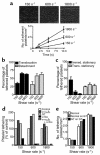
Figure 1
Figure 1
Dynamics of platelet aggregation under flow. Fluorescently labeled human platelets in whole blood were perfused over confluent spread platelet monolayers at 150, 600, and 1800 s–1 for 1 minute. (a) The images depict the shear-dependent increase in platelet adhesion after 1 minute of flow. The number of platelets tethering over a 10-second time period is demonstrated in the line graph. Each tethered platelet was examined over a further 10-second time period, and the percentage of (b) platelets translocating and/or detaching and/or (c) forming stationary (immediate stationary or translocating then stationary) contacts was determined. Results represent mean ± SEM (n = 4). (d) Role of GPIbα in mediating platelet–platelet interactions under flow. Before perfusion, blood was incubated for 10 minutes with buffer (control), anti-αIIbβ3 (α-αIIbβ3), anti-GPIbα (α-Ibα), or both α-αIIbβ3 and Ibα antibodies. In other studies GT platelets were perfused over normal platelet monolayers. (e) Fluorescently labeled platelets from a normal donor or a patient with GT were perfused over platelet monolayers prepared from a normal (normal) or a GT donor (normal on GT; GT on GT). The level of platelet tethering in the first 5 seconds of perfusion was determined. These results are representative of 3 experiments and are indicative of the tethering process over the full 1-minute perfusion period.
Figure 2
Figure 2
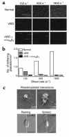
Role of platelet vWf in mediating platelet tethering under flow. (a) Fluorescently labeled human platelets in whole blood from a normal donor were perfused over a platelet monolayer prepared from a normal donor (normal) or, alternatively, whole blood from an individual with vWD was perfused over a vWf-deficient platelet monolayer (vWD), in the presence or absence of an anti-αIIbβ3 (α-αIIbβ3) antibody at the indicated shear rates. (b) Quantitation of platelet tethering in the first 5 seconds of perfusion. These results are representative of 5 experiments and are indicative of the tethering process over the full 1-minute perfusion period. (c) Platelet–platelet interactions occur at the site of vWf localization. Whole blood was perfused over a spread platelet monolayer at 600 s–1 for 2 minutes. Upper panels: These scanning electron micrographs demonstrate platelet tethering onto the granulomere of spread platelets (left image) and the early formation of a platelet aggregate (right image). These images are representative of over 100 cells examined. Lower panels: The images with white immunofluorescence (α-vWf; right half) partially overlaid onto the DIC image (left half) demonstrate that vWf becomes expressed on the surface of activated (spread) platelets and is specifically localized to the granulomere (right image). Note in the left image that vWf is not present on the surface of resting platelets. Bar, 5 μm.
Figure 3
Figure 3
Relative roles of plasma and platelet vWf in initiating platelet aggregation under flow. Fluorescently labeled whole blood from a normal donor (normal blood) or a vWD patient (vWD blood) was perfused over pre-formed monolayers prepared with platelets from a normal donor (normal monolayer) or from a vWD patient (vWD monolayer) at the indicated shear rates. The level of platelet tethering in the first 5 seconds of flow (a) and the 5 seconds of flow after 30 seconds of perfusion (b) was determined. Results are presented as mean ± SEM (n = 5). Note: The reduction in number of tethering platelets between 30–35 seconds relative to 0–5 seconds reflects a decrease in the number of reactive sites available for platelet capture as a consequence of increased platelet adhesion. (c and d) Role of platelet vWf in promoting thrombus growth at high shear. Fluorescently labeled whole blood from a normal donor or from a vWD patient was perfused over collagen at 150 s–1 for 2 minutes. This was followed by immediate perfusion of normal blood (normal), vWD blood (vWD), or vWD blood supplemented with purified soluble vWf (10 μg/mL; vWD + vWf) over the pre-formed reactive platelet surface at 1800 s–1. Thrombus growth was monitored in real time by confocal microscopy at 1-minute intervals for 5 minutes. (c) Platelet thrombi formed after 5 minutes of perfusion were reconstructed using a computer-assisted image-analysis program. The upper panels represent an oblique view to demonstrate surface coverage while the lower panels represent a side-on view to demonstrate differences in thrombus height. (d) Total thrombus volume in a field of interest (25,192 μm2) was calculated using Image Tool. Note: When calculating thrombus volumes, the first 2 μm were omitted from analysis to exclude the pre-formed platelet monolayers. The results are from 1 experiment representative of 2.
Figure 4
Figure 4
Dynamics of rat platelet aggregation in vitro and in vivo. Fluorescently labeled rat platelets in whole blood were perfused over spread rat platelet monolayers at 150, 600, and 1800 s–1 for 1 minute. (a) The percentage of tethered platelets translocating and/or detaching over a 10-second period is demonstrated. (b) Before perfusion, blood was incubated for 10 minutes with buffer (control), anti-αIIbβ3 (α-αIIbβ3), anti-GPV (α-GPV), or anti-GPIbα (α-Ibα) antibody fragments. In these studies the number of platelets tethering over a 5-second time period is demonstrated. Results represent mean ± SEM (n = 4). (c) Dynamics of platelet aggregation in vivo. Vascular injury and thrombus formation in rat mesenteric vessels was induced by photoactivation of Rose Bengal. The percentage of platelets translocating and/or detaching from the surface of arterial and venous thrombi was examined over a 10-second time period using DIC microscopy. (d) Following thrombus formation, fluorescently labeled, washed rat platelets, preincubated with buffer (control), anti-GPIbα (α-GPIbα), anti-αIIbβ3 (α-αIIbβ3), or anti-GPV (α-GPV) antibody fragments were injected intravenously. Pre-formed platelet thrombi (white arrowheads) were viewed by differential interference contrast (upper panels; DIC) whereas tethering of injected platelets to the surface of pre-formed thrombi was observed by fluorescence microscopy (lower panels; ×63). For clarity, the mesenteric arteriole walls have been demarcated using continuous lines, whereas the surface of thrombi have been outlined using discontinuous lines. Uninjured refers to a mesenteric vessel that has not been subjected to photo-induced injury. These images are from 5 experiments and are representative of more than 50 independent experiments. Note the α-αIIbβ3–treated platelets that adhered to the surface of arterial thrombi formed transient tethers. All these cells subsequently translocated and/or detached from the thrombus.
Figure 5
Figure 5
A revised model of platelet aggregation. (a and b) The mechanisms by which platelets in suspension aggregate following exposure to a soluble agonist or high shear stress are depicted. (c) The multistep mechanism by which circulating platelets form stationary adhesion contacts with the luminal surface of a growing thrombus under physiological flow conditions is demonstrated. (a) The addition of a soluble agonist to a stirred platelet suspension leads to the activation of integrin αIIbβ3 (GPIIb-IIIa) and its subsequent binding to soluble fibrinogen. Fibrinogen’s dimeric structure enables it to cross-link adjacent activated platelets and mediate stable platelet aggregation (5, 38). (b) Under high shear conditions, vWf becomes the relevant ligand responsible for platelet activation. Shear-induced binding of soluble vWf to GPIb-V-IX induces platelet activation. At elevated levels of shear, stable platelet aggregation is thought to be dependent primarily on vWf binding to GPIIb-IIIa (7, 39). (c) Upon vascular injury, platelets are recruited to the subendothelium where they become activated, releasing their granule contents. vWf expressed on the surface of immobilized platelets tethers circulating platelets by binding GPIbα. The majority of platelets that tether subsequently translocate or detach from the surface of immobilized platelets. The formation of stationary adhesion contacts is dependent on the cross-linking of integrin αIIbβ3 on the surface of free-flowing and immobilized platelets by plasma vWf or fibrinogen. This multistep adhesion process operates at both arterial and venous levels of shear and appears to be the predominant mechanism by which platelets aggregate in vivo. However, note that at venous levels of shear, platelets can also form immediate stationary adhesion contacts with immobilized platelets independent of the vWf-GPIbα interaction.

Comment in

References

    1. Savage B, Almus-Jacobs F, Ruggeri ZM. Specific synergy of multiple substrate-receptor interactions in platelet thrombus formation under flow. Cell. 1998;94:657–666. - PubMed
    1. Tschopp TB, Weiss HJ, Baumgartner HR. Decreased adhesion of platelet to subendothelium in von Willebrand’s disease. J Lab Clin Med. 1973;83:296–300. - PubMed
    1. Weiss HJ, Turitto VT, Baumgartner HR. Effect of shear rate on platelet interaction with subendothelium in citrated and native blood. I. Shear rate-dependent decrease of adhesion in von Willebrand’s disease and the Bernard-Soulier syndrome. J Lab Clin Med. 1978;92:750–764. - PubMed
    1. Savage B, Saldivar E, Ruggeri ZM. Initiation of platelet adhesion by arrest onto fibrinogen and translocation on von Willebrand factor. Cell. 1996;84:289–297. - PubMed
    1. Holmsen, H. 1986. Platelet responses and metabolism. CRC Press. Boca Raton, FL. pp. 11.

Publication types

MeSH terms

Substances