. 2000 Mar;105(6):R9-R14.
doi: 10.1172/JCI9051.

Mature dendritic cells boost functionally superior CD8(+) T-cell in humans without foreign helper epitopes

Affiliations

Mature dendritic cells boost functionally superior CD8(+) T-cell in humans without foreign helper epitopes

M V Dhodapkar et al. J Clin Invest. 2000 Mar.

Abstract

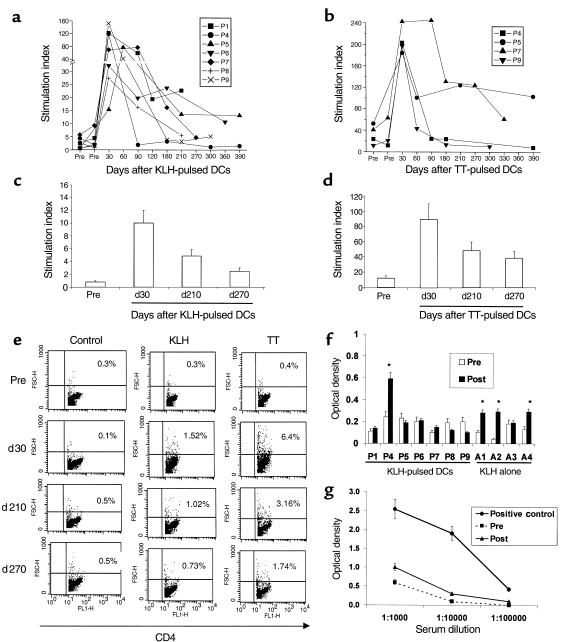
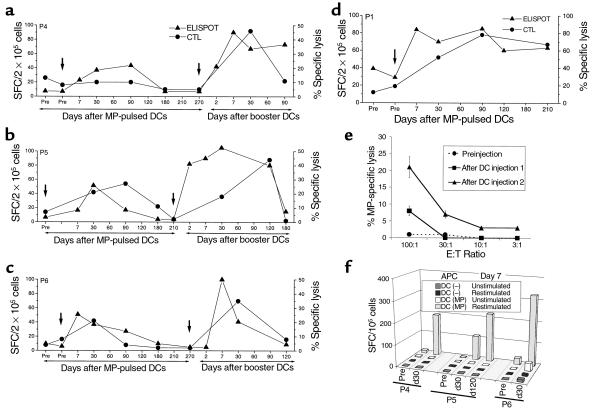
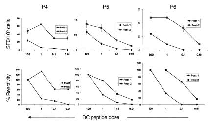
We have recently shown that a single injection of mature, antigen-pulsed, human dendritic cells (DCs) rapidly elicits CD4(+) and CD8(+) T-cell immunity in vivo. The DCs were pulsed with 2 foreign proteins, keyhole limpet hemocyanin (KLH) and tetanus toxoid (TT), as well as an HLA A2.1-restricted influenza matrix peptide (MP). Responses to all 3 antigens peaked at 30-90 days after immunization and declined thereafter. To determine if the foreign helper proteins (TT and KLH) were essential for CD8(+) T-cell responses to the viral peptide, we reinjected 3 of the HLA-2.1 subjects with mature DCs pulsed with MP alone. All 3 volunteers showed a rapid boost in MP-specific immunity, and freshly sampled blood from 1 contained cytolytic T cells. In all 3 subjects, CD8(+) T-cell responses to booster DCs were faster and of greater magnitude than the responses to the first DC injection. Importantly, the T cells that proliferated after booster DC treatment secreted interferon-gamma upon challenge with much lower doses of viral peptide than those elicited after the first injection, indicating a higher functional avidity for the ligand. These data begin to outline the kinetics of T-cell immunity in response to DCs and demonstrate that booster injections of mature DCs enhance both qualitative and quantitative aspects of CD8(+) T-cell function in humans.


Figures

Figure 1
Figure 1
TT- and KLH-specific immunity after DC injection. (a) Longevity of KLH-specific immune response after single DC injection. For each measurement shown, 105 freshly isolated PBMCs were incubated in the presence or absence of 10 μg/mL KLH for 5 days, and proliferation was measured as 3H-TdR incorporation. Results are expressed as stimulation index. SEM < 30%; cpm without antigen < 5 × 10³. (b) Longevity of TT-specific immune response after single DC injection. For each measurement shown, 105 freshly isolated PBMCs were incubated in the presence or absence of 3 μg/mL TT for 5 days, and proliferation was measured as 3H-TdR incorporation. Results are expressed as stimulation index. SEM < 30%. (c) Evaluation of longevity of KLH-specific response using cryopreserved specimens. Pre- and postimmunization specimens were thawed together and cultured in the presence or absence of 10 μg/mL KLH for 5 days. Antigen-specific proliferation was measured as 3H-TdR incorporation. Results are shown as stimulation index. Data shown are for 1 subject (P5), representative of 3 subjects tested. (d) Evaluation of longevity of TT- specific response using cryopreserved specimens. Pre- and postimmunization specimens were thawed together and cultured in the presence or absence of 3 μg/mL TT for 5 days. Antigen-specific proliferation was measured as 3H-TdR incorporation. Results are shown as stimulation index. Data shown are for 1 subject (P5), representative of 2 subjects tested. (e) Quantification of CD4 proliferative response as percent of CD4 blasts. T-cell cultures from the experiment in c and d were stained for CD4 and analyzed by flow cytometry. Percent of CD4 blasts were quantified as percent of CD4+ T cells with high forward scatter, noted in the upper-right quadrant. Data shown are gated for CD4+ cells. (f and g) Detection of KLH-specific antibodies. (f) Presence of KLH-specific antibodies was determined in sera before and 3 months after immunization by ELISA as described in Methods. Data shown are at a serum dilution of 1:104. Because of variable background, reactivity was assayed by comparing pre- and postimmunization samples in the same individual. All assays were repeated to verify results. *P < 0.05. (g) Representative data from a subject (A1) injected with KLH alone. KLH-specific antibodies were detected in only 1 of 7 volunteers (P4) injected with KLH-pulsed DCs. Positive control was an individual with known high-titer anti-KLH antibodies.
Figure 2
Figure 2
Enhancement of antigen-specific CD8+ T cells in vivo. (ad) Kinetics of antigen-specific CD8+ T cells following DC injection(s). MP-specific interferon-γ–producing T cells in freshly isolated uncultured T cells were quantified using an ELISPOT assay (triangles). Results are shown as the number of spot-forming cells (SFC)/2 × 105 PBMC. MP-specific CTLs were quantified after 7-day coculture with MP-pulsed mature DCs (DC/T cell ratio 30:1). Data shown are percent of MP-specific lysis at each time point (effector/target [E/T] ratio 20:1) after subtracting lysis with unpulsed T2 targets and that using unpulsed DCs (circles). SEM < 25%. Arrows indicate the timing of DC injections. The subjects were: a, P4; b, P5; c, P6; and d, P1. (e) Detection of circulating lytic effectors after booster DC injection. Freshly isolated bulk PBMCs of a study subject (P4) from before injection and 7 days after booster DC injections 1 and 2 were directly tested for the presence of MP-specific lytic effectors using MP-pulsed T2 cells as targets at varying E/T ratios. Data shown are percent of MP-specific lysis after subtracting lysis of unpulsed T2 cells. (f) Enhancement of MP-specific IFN-γ–producing T cells after booster DC injection, recall ELISPOT assay. Cryopreserved PBMCs from before (pre) and 30 or 120 days after booster DC injection were thawed together and cocultured for 7 days with freshly generated mature DCs (DC/PBMC ratio 1:30), either unpulsed, DC(–), or pulsed with 1 μg/mL MP, DC (MP), without exogenous cytokines. On day 7, T cells were either left unstimulated or restimulated by the addition of 10 μg/mL MP peptide and transferred to an ELISPOT plate. MP-specific IFN-γ–producing T cells were quantified using a 16-hour ELISPOT assay. SEM < 30%.
Figure 3
Figure 3
MP-specific T cells after booster DCs show greater peptide sensitivity than those after the first injection. Cryopreserved PBMCs from 30 days after the first (post-1; circles) and second booster antigen-pulsed (post-2; squares) DC injection were thawed together and cocultured with freshly generated mature DCs (DC/PBMC ratio 1:30), pulsed with graded (0.01–100 ng/mL) MP. After overnight culture, the presence of MP-specific T cells (expressed as SFCs) was quantified using an ELISPOT. (Top) Effect of peptide dose pulsed on DCs on the detection of MP-specific IFN-γ–producing cells. (Bottom) Data expressed as percent of reactivity seen with DCs pulsed with the highest dose of MP (100 ng/mL).

Comment in

References

    1. Raychaudhuri S, Rock KL. Fully mobilizing host defense: building better vaccines. Nat Biotechnol. 1998;16:1025–1031. - PubMed
    1. Banchereau J, Steinman RM. Dendritic cells and the control of immunity. Nature. 1998;392:245–252. - PubMed
    1. Dhodapkar MV, et al. Rapid generation of broad T-cell immunity in humans after a single injection of mature dendritic cells. J Clin Invest. 1999;104:173–180. - PMC - PubMed
    1. Murphy G, Tjoa B, Ragde H, Kenny G, Boynton A. Phase I clinical trial: T-cell therapy for prostate cancer using autologous dendritic cells pulsed with HLA-A020-specific peptides from prostate-specific peptides from prostate-specific membrane antigen. Prostate. 1996;29:371–380. - PubMed
    1. Nestle FO, et al. Vaccination of melanoma patients with peptide- or tumor lysate-pulsed dendritic cells. Nat Med. 1998;4:328–332. - PubMed

Publication types