. 2000 Mar 20;191(6):937-48.
doi: 10.1084/jem.191.6.937.

Self-recognition of CD1 by gamma/delta T cells: implications for innate immunity

Affiliations

Self-recognition of CD1 by gamma/delta T cells: implications for innate immunity

F M Spada et al. J Exp Med. .

Abstract

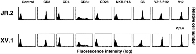
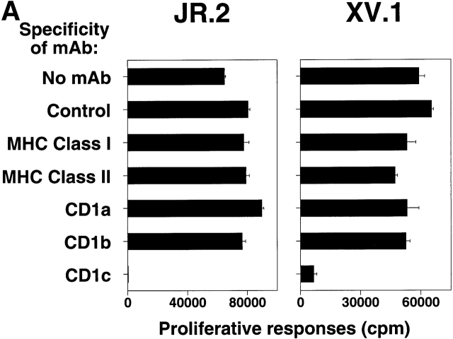
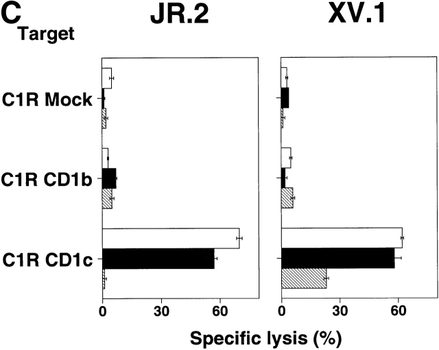
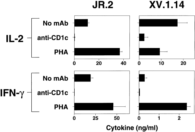
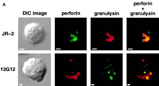
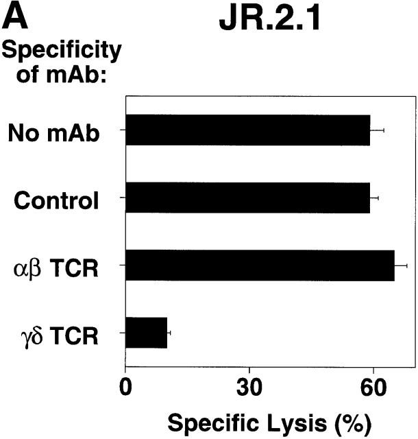
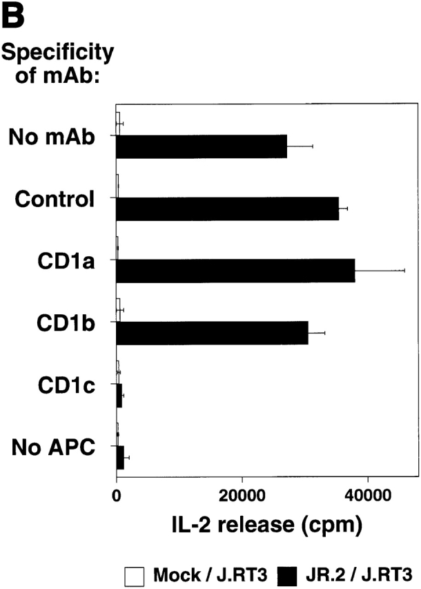
The specificity of immunoglobulins and alpha/beta T cell receptors (TCRs) provides a framework for the molecular basis of antigen recognition. Yet, evolution has preserved a separate lineage of gamma/delta antigen receptors that share characteristics of both immunoglobulins and alpha/beta TCRs but whose antigens remain poorly understood. We now show that T cells of the major tissue gamma/delta T cell subset recognize nonpolymorphic CD1c molecules. These T cells proliferated in response to CD1+ presenter cells, lysed CD1c+ targets, and released T helper type 1 (Th1) cytokines. The CD1c-reactive gamma/delta T cells were cytotoxic and used both perforin- and Fas-mediated cytotoxicity. Moreover, they produced granulysin, an important antimicrobial protein. Recognition of CD1c was TCR mediated, as recognition was transferred by transfection of the gamma/delta TCR. Importantly, all CD1c-reactive gamma/delta T cells express V delta 1 TCRs, the TCR expressed by most tissue gamma/delta T cells. Recognition by this tissue pool of gamma/delta T cells provides the human immune system with the capacity to respond rapidly to nonpolymorphic molecules on professional antigen presenting cells (APCs) in the absence of foreign antigens that may activate or eliminate the APCs. The presence of bactericidal granulysin suggests these cells may directly mediate host defense even before foreign antigen-specific T cells have differentiated.


Figures

Figure 1
Figure 1
Flow cytometric analysis of JR.2 and XV.1 lines. The mAbs used were P3 (IgG control), SPV-T3b (anti-CD3), OKT4 (anti-CD4), OKT8 (anti-CD8α), 9.3 (anti-CD28), DX1 (anti–NKR-P1A), anti–TCR-δ1 (pan anti-Cδ TCR), δTCS1 (anti-Vδ1/Jδ1), TiγA (anti-Vγ2), and 4A11 (anti-Vγ1.4).
Figure 2
Figure 2
CD1c-specific recognition by JR.2 and XV.1 γ/δ T cell lines. (A) The proliferative response of JR.2 and XV.1 lines to CD1c+ dendritic cells was inhibited by anti-CD1c mAb (F10/2A3) but not by anti–MHC class I, anti–MHC class II, anti-CD1a, and anti-CD1b mAbs. (B) The JR.2 T cell line proliferated in response to HeLa CD1c cells but not to HeLa Mock cells. Background proliferation of HeLa Mock and HeLa CD1c cells alone was 872 and 1,197 cpm, respectively. (C) C1R Mock, C1R CD1b, and C1R CD1c targets were 51Cr labeled and tested in cytolytic assays with JR.2 and XV.1 T cell lines at different E/T ratios (30:1 is shown) in the presence of anti-CD1b (BCD1b3.2) or anti-CD1c (F10/213) mAbs. Note that JR.2 and XV.1 lysed only C1R CD1c transfectant cells and that lysis was blocked by anti-CD1c mAb. White bars, no mAb; black bars, anti-CD1b; gray bars, anti-CD1c.
Figure 2
Figure 2
CD1c-specific recognition by JR.2 and XV.1 γ/δ T cell lines. (A) The proliferative response of JR.2 and XV.1 lines to CD1c+ dendritic cells was inhibited by anti-CD1c mAb (F10/2A3) but not by anti–MHC class I, anti–MHC class II, anti-CD1a, and anti-CD1b mAbs. (B) The JR.2 T cell line proliferated in response to HeLa CD1c cells but not to HeLa Mock cells. Background proliferation of HeLa Mock and HeLa CD1c cells alone was 872 and 1,197 cpm, respectively. (C) C1R Mock, C1R CD1b, and C1R CD1c targets were 51Cr labeled and tested in cytolytic assays with JR.2 and XV.1 T cell lines at different E/T ratios (30:1 is shown) in the presence of anti-CD1b (BCD1b3.2) or anti-CD1c (F10/213) mAbs. Note that JR.2 and XV.1 lysed only C1R CD1c transfectant cells and that lysis was blocked by anti-CD1c mAb. White bars, no mAb; black bars, anti-CD1b; gray bars, anti-CD1c.
Figure 3
Figure 3
JR.2 and XV.1 γ/δ T cell lines show a Th1-like cytokines profile. JR.2.1 and XV.1.14 clones were incubated with CD1c+ dendritic cells for 24 and 48 h. The culture supernatants were collected and assayed for IL-2, IFN-γ, IL-4, and IL-10. Note that the JR.2.1 and XV.1.14 clones produced IFN-γ and IL-2 in response to exposure to CD1+ dendritic cells. IL-4 and IL-10 were not detected (data not shown).
Figure 5
Figure 5
Expression of granulysin by γ/δ T cells. (A) Colocalization of granulysin and perforin in the cytolytic granules. Differential interference contrast images of γ/δ T cells are shown on the left. Confocal microscopic analysis of fluorescent immunostaining of perforin and granulysin of the JR.2 CD1c-reactive γ/δ T cell line and 12G12 phosphoantigen–specific γ/δ T cell clone are shown as labeled. Superimposed figures are showed in the right panels. Note that the counterstaining were exchanged such that perforin is green in the top panel but red in the bottom panel, and granulysin is red in the top panel but green in the bottom panel. (B) Localization of granulysin to the cytotoxic granules of γ/δ T cells by immunoelectron microscopy. The presence of granulysin in the cytotoxic granules of JR.2 (bottom) and 12G12 (top) is demonstrated by the gold labeling (small particles) seen with antigranulysin mAb staining. c, centriole; e, endosome; P, plasma membrane; m, mitochondria; n, nucleus; G, Golgi apparatus. Bar, 200 nm.
Figure 5
Figure 5
Expression of granulysin by γ/δ T cells. (A) Colocalization of granulysin and perforin in the cytolytic granules. Differential interference contrast images of γ/δ T cells are shown on the left. Confocal microscopic analysis of fluorescent immunostaining of perforin and granulysin of the JR.2 CD1c-reactive γ/δ T cell line and 12G12 phosphoantigen–specific γ/δ T cell clone are shown as labeled. Superimposed figures are showed in the right panels. Note that the counterstaining were exchanged such that perforin is green in the top panel but red in the bottom panel, and granulysin is red in the top panel but green in the bottom panel. (B) Localization of granulysin to the cytotoxic granules of γ/δ T cells by immunoelectron microscopy. The presence of granulysin in the cytotoxic granules of JR.2 (bottom) and 12G12 (top) is demonstrated by the gold labeling (small particles) seen with antigranulysin mAb staining. c, centriole; e, endosome; P, plasma membrane; m, mitochondria; n, nucleus; G, Golgi apparatus. Bar, 200 nm.
Figure 4
Figure 4
Analysis of cytotoxicity mechanisms used by CD1c-reactive and prenyl pyrophosphate–specific γ/δ T cells. CD1c-reactive Vγ2/Vδ1 T cells, JR.2 and IDP2, and prenyl pyrophosphate antigen–specific Vγ2/Vδ2 T cell clones, 12G12, HD.108, CP.1.15, and DG.SF68, were used in cytolytic assays performed in the presence or absence of anti-Fas mAb to block Fas-mediated cytolysis or after treatment with strontium ions to block perforin-mediated cytolysis. The mycolic acid–specific, CD1b-restricted, CD4CD8 α/β T cell line, DN1, is shown as a control. MEP is an alkyl phosphate analogue of the IPP antigen recognized by Vγ2/Vδ2 T cells and was used to stimulate the prenyl pyrophosphate–specific clones. The E/T ratio was 10:1. Targets for CD1c-reactive and prenyl pyrophosphate–specific γ/δ T cells were C1R CD1c cells. Targets for the CD1b-restricted clone were C1R CD1b cells that had been incubated for 16 h at 37°C with M. tuberculosis sonicate at 1 μg/ml. Note that the cytolytic activity mediated by γ/δ T cells was blocked by both treatments and thus depends on both granule secretion and Fas–FasL interactions.
Figure 7
Figure 7
Diverse CDR3 junction region of two CD1c-reactive Vγ2/Vδ1 T cell clones, showing nucleotide sequence analysis of the γ and the δ gene rearrangements of JR.2 CD1c-reactive clone. Deduced amino acid sequences are in the single letter code. IDP2 sequence data are taken from Hata et al. (reference 64) and Krangel et al. (reference 65) and are included for comparison.
Figure 6
Figure 6
TCR-γ/δ mediates CD1c recognition. (A) JR.2.1 T cell clone lysed C1R CD1c cells. Lysis was blocked by anti-CD1c mAb (F10/2A3; not shown) as well by anti-Cδ TCR mAb (anti–TCR-δ1). E/T 30:1. (B) JR.2 Vγ2/Vδ1 transfectant recognizes CD1c. The TCR J.RT3-T3 cell line was transfected with cDNAs encoding the Vγ2/Vδ1 TCR from the JR.2 cell line or was mock transfected. The resulting JR.2/J.RT3 or Mock/J.RT3 cell lines were cultured with CD1+ dendritic cells in the presence or absence of mAbs to CD1a, CD1b, and CD1c, and the supernatants were harvested after 24 h. IL-2 release was assessed by the proliferation of the IL-2–dependent HT-2 T cell line. Note that the JR.2/J.RT3 but not the Mock/J.RT3 cell line released IL-2 when cultured with CD1+ monocyte-derived dendritic cells. This IL-2 release was completely blocked by the addition of an anti-CD1c mAb.
Figure 6
Figure 6
TCR-γ/δ mediates CD1c recognition. (A) JR.2.1 T cell clone lysed C1R CD1c cells. Lysis was blocked by anti-CD1c mAb (F10/2A3; not shown) as well by anti-Cδ TCR mAb (anti–TCR-δ1). E/T 30:1. (B) JR.2 Vγ2/Vδ1 transfectant recognizes CD1c. The TCR J.RT3-T3 cell line was transfected with cDNAs encoding the Vγ2/Vδ1 TCR from the JR.2 cell line or was mock transfected. The resulting JR.2/J.RT3 or Mock/J.RT3 cell lines were cultured with CD1+ dendritic cells in the presence or absence of mAbs to CD1a, CD1b, and CD1c, and the supernatants were harvested after 24 h. IL-2 release was assessed by the proliferation of the IL-2–dependent HT-2 T cell line. Note that the JR.2/J.RT3 but not the Mock/J.RT3 cell line released IL-2 when cultured with CD1+ monocyte-derived dendritic cells. This IL-2 release was completely blocked by the addition of an anti-CD1c mAb.

Comment in

References

    1. Hiromatsu K., Yoshikai Y., Matsuzaki G., Ohga S., Muramori K., Matsumoto K., Bluestone J.A., Nomoto K. A protective role of γ/δ T cells in primary infection with Listeria monocytogenes in mice. J. Exp. Med. 1992;175:49–56. - PMC - PubMed
    1. Ladel C.H., Blum C., Dreher A., Reifenberg K., Kaufmann S.H.E. Protective role of γ/δ T cells and α/β T cells in tuberculosis. Eur. J. Immunol. 1995;25:2877–2881. - PubMed
    1. Ladel C.H., Hess J., Daugelat S., Mombaerts P., Tonegawa S., Kaufmann S.H.E. Contribution of α/β and γ/δ T lymphocytes to immunity against Mycobacterium bovis bacillus Calmette Guérinstudies with T cell receptor-deficient mutant mice. Eur. J. Immunol. 1995;25:838–846. - PubMed
    1. D'Souza C.D., Cooper A.M., Frank A.A., Mazzaccaro R.J., Bloom B.R., Orme I.M. An anti-inflammatory role for γδ T lymphocytes in acquired immunity to Mycobacterium tuberculosis . J. Immunol. 1997;158:1217–1221. - PubMed
    1. Tsuji M., Mombaerts P., Lefrancois L., Nussenzweig R.S., Zavala F., Tonegawa S. γδ T cells contribute to immunity against the liver stages of malaria in αβ T-cell-deficient mice. Proc. Natl. Acad. Sci. USA. 1994;91:345–349. - PMC - PubMed

Publication types

MeSH terms