. 2000 Mar 20;191(6):1005-16.
doi: 10.1084/jem.191.6.1005.

Deregulated E2F transcriptional activity in autonomously growing melanoma cells

Affiliations

Deregulated E2F transcriptional activity in autonomously growing melanoma cells

R Halaban et al. J Exp Med. .

Abstract

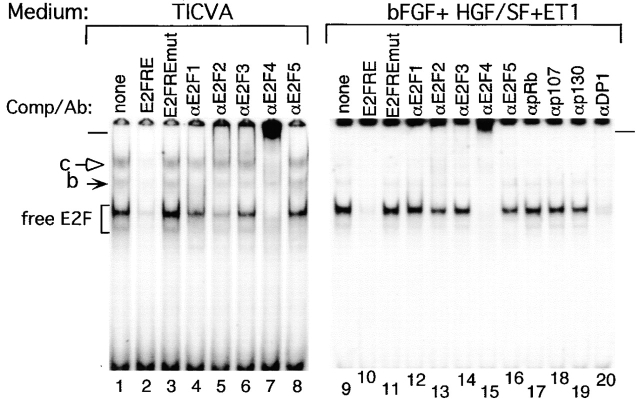
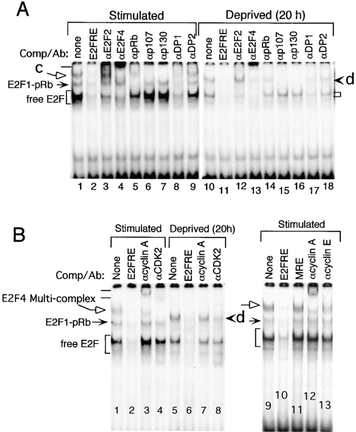
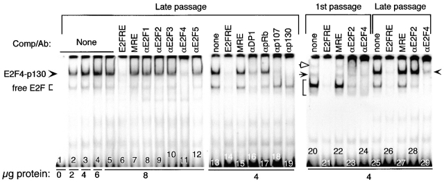
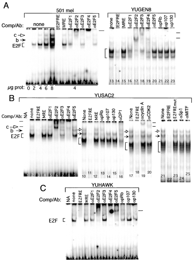
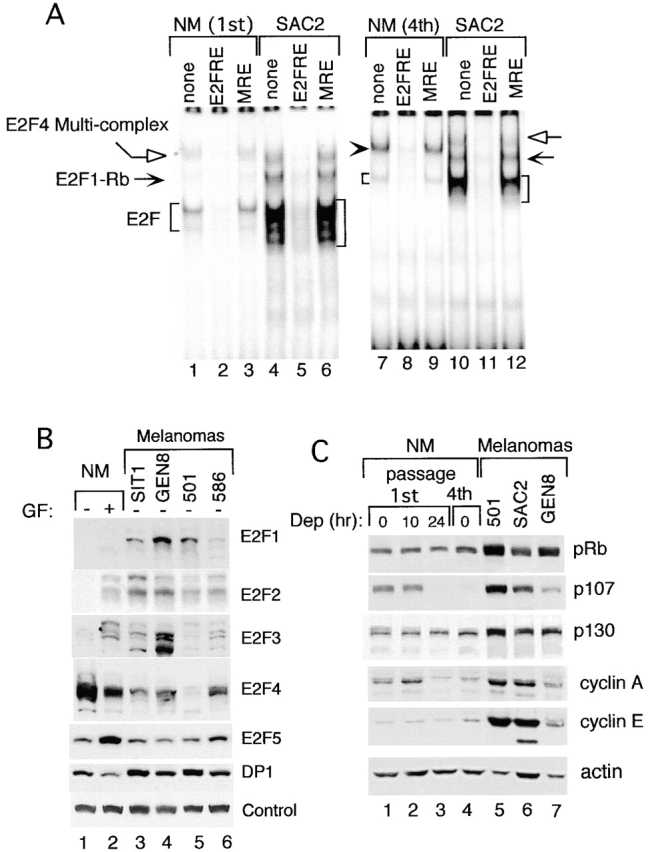
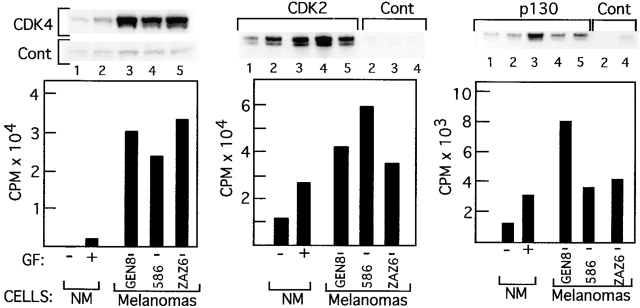
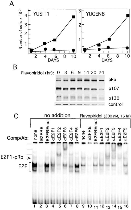
Inactivation of the retinoblastoma tumor suppressor protein (pRb) has been implicated in melanoma cells, but the molecular basis for this phenotype has not yet been elucidated, and the status of additional family members (p107 and p130, together termed pocket proteins) or the consequences on downstream targets such as E2F transcription factors are not known. Because cell cycle progression is dependent on the transcriptional activity of E2F family members (E2F1-E2F6), most of them regulated by suppressive association with pocket proteins, we characterized E2F-pocket protein DNA binding activity in normal versus malignant human melanocytes. By gel shift analysis, we show that in mitogen-dependent normal melanocytes, external growth factors tightly controlled the levels of growth-promoting free E2F DNA binding activity, composed largely of E2F2 and E2F4, and the growth-suppressive E2F4-p130 complexes. In contrast, in melanoma cells, free E2F DNA binding activity (E2F2 and E2F4, to a lesser extent E2F1, E2F3, and occasionally E2F5), was constitutively maintained at high levels independently of external melanocyte mitogens. E2F1 was the only family member more abundant in the melanoma cells compared with normal melanocytes, and the approximately fivefold increase in DNA binding activity could be accounted for mostly by a similar increase in the levels of the dimerization partner DP1. The continuous high expression of cyclin D1, A2, and E, the persistent cyclin-dependent kinase 4 (CDK4) and CDK2 activities, and the presence of hyperphosphorylated forms of pRb, p107, and p130, suggest that melanoma cells acquired the capacity for autonomous growth through inactivation of all three pocket proteins and release of E2F activity, otherwise tightly regulated in normal melanocytes by external growth factors.


Figures

Figure 1
Figure 1
Growth factor regulation of E2F DNA binding activity and expression of growth-regulatory proteins in normal human melanocytes. Melanocytes (first passage) were used in parallel to assay for proliferative responses (A); E2F DNA binding activity (B); and pocket protein and cyclin expression (C). (A, a) Melanocytes grown in TICVA/supplemented basal medium were seeded in 24-well plates (1.4 × 10⁵ melanocytes/well) in either TICVA-supplemented (0) or deprived medium for the indicated period of time. [3H]Thymidine incorporation was for 1 h at different time points as indicated. Data are averages of triplicate wells. SE did not exceed 10%. (A, b) Melanocytes (0.7 × 10⁵) were seeded into 48-well plates and exposed to OptiMEM medium with or without peptide growth factors. [3H]Thymidine was added 24 h after the addition of experimental medium for 4 h. Data are averages of triplicate wells normalized to 1 h/1.4 × 105 melanocytes, as in b. Growth factors included: bFGF/FGF2 (H, 10 ng/ml), HGF/SF (F, 100 ng/ml), ET-1 (E, 0.1 μM), or combinations thereof. NA, no additions. SD did not exceed 13% of total counts. (B) EMSA of whole cell lysates prepared from melanocytes grown in TICVA/serum-supplemented medium and harvested at the exponential phase of growth (lanes 1–3), or after TICVA deprivation for the time indicated (lanes 4–8), as in A, a. Alternatively, melanocytes were incubated with peptide growth factors as in A, b, and harvested 24 h later (lanes 9 and 10). E2F EMSA reactions contained 4 μg of total cellular proteins and an end-labeled DNA fragment derived from the DHFR promoter. Excess (10 ng) unlabeled double-stranded oligonucleotide containing wild-type E2F responsive element site (E2FRE) or mutant E2FRE (E2FREmut) were added to gel shift assays where indicated. GF, growth factor; additional abbreviations as indicated in the legend to A, b. (C) Western blot analyses of pocket proteins and cyclins of melanocytes treated as in A, a (lanes 1–6) and A, b (lanes 7–14) with antibodies as indicated. Equal loading of the 6% gel used to fractionate pocket proteins is indicated by the spurious band (control), whereas equal loading of the 10% used to fractionate the cyclins is represented by actin staining (actin).
Figure 2
Figure 2
Identification of the components in the E2F DNA binding activity in nuclear extracts. Gel shift analyses of melanocytes grown in TICVA-supplemented basal medium (lanes 1–8), or in OptiMEM supplemented with 2% FBS and peptide growth factors bFGF/FGF2, HGF/SF, and ET-1, for 24 h. E2F EMSA reactions contained 4 μg of nuclear proteins and an end-labeled DNA fragment as described in the legend to Fig. 1. Antibodies specific for each of the E2F family members, E2F1–E2F5, or each of the pocket proteins, pRb, p107, or p130, or the E2F dimerization partner, DP1, were added to the binding reactions as indicated to identify proteins in specific complexes. Bracket marks free E2F species, solid and open arrows (marked b and c, respectively) point at complex E2F binding activity, and bars on the left and right of the figure mark the positions of the supershifts.
Figure 3
Figure 3
Gel shift analyses of nuclear extracts from stimulated versus growth-arrested melanocytes. (A and B) Melanocytes grown in TICVA plus serum-supplemented medium were harvested at the exponential phase of growth (Stimulated), or after transfer to basal medium without TICVA, for 20 h (Deprived [20 h]). E2F EMSA reactions contained end-labeled DNA fragment derived from the DHFR promoter, 4 μg of nuclear proteins, cold competitors, or antibodies, where indicated and as described above. Free E2F binding activity is marked by brackets (free E2F), and complex activities by solid (E2F1-pRb) and open arrows (c, or E2F4 Multi-complex), and arrowhead (d). Antibodies to specific proteins were added to the reaction mixtures as indicated. Bars mark the supershifts.
Figure 4
Figure 4
A shift to growth-arrest pattern of E2F DNA binding activity in senescing normal melanocytes. Nuclear extracts were prepared from fourth (lanes 2–19, 25–29) or first passage melanocyte cultures grown in TICVA-supplemented basal medium and harvested at the exponential growth phase. DNA binding reactions contained 0–8 μg of nuclear proteins (as indicated) and an end-labeled DNA fragment derived from the DHFR promoter as described above. The reaction mixtures contained excess (10 ng) double-stranded oligonucleotide encoding wild-type E2F site (E2FRE), the unrelated MRE site, or specific antibodies as indicated. Brackets and arrowheads point at the positions of the free E2F and E2F4-p130 suppressive complexes, respectively. The position of E2F1–pRb and the multimeric E2F4–p130 (p107)/cyclin A–CDK2 are indicated by solid and open arrows, respectively.
Figure 5
Figure 5
Free E2F DNA binding activity is abundant in melanoma cells and tumors. Nuclear extracts (A, 0–8 μg/assay; B and C, 4 μg/assay) from metastatic melanoma cells (501 mel, YUGEN8, and YUSAC2) or a freshly isolated melanoma tumor (YUHAWK) were used in EMSAs as described above. All cells in culture were from the exponential growth phase. Excess unlabeled competitors or specific antibody were added to the reaction mixtures as described above. Complexes are marked as above. NA, reaction contained radioactive E2FRE without cell extracts or any other additions.
Figure 6
Figure 6
Upregulation of E2F DNA binding activity in proliferating melanoma cells compared with normal melanocytes as a function of pocket protein and E2F expression. (A) EMSA showing the relative amounts of E2F DNA binding activity in normal melanocytes (NM, first and fourth passage) compared with melanoma cells YUSAC2 (SAC2), all harvested at the exponential growth phase. Each reaction mixture contained 4 μg nuclear proteins, radiolabeled probe, and/or competitors (where indicated) as described above. Normal melanocytes were grown in TICVA-supplemented basal medium, whereas melanoma cells YUSAC2 were grown in basal medium without TICVA. (B) Expression of E2F1–E2F5 and DP1 in normal melanocytes compared with melanoma cells. Shown are Western blots with the respective antibodies (as marked) using nuclear extracts (prepared as described for gel shift analysis) harvested from exponentially growing (+) or 24 h TICVA–deprived normal melanocytes (NM), or from proliferating melanoma cells YUSIT1 (SIT1), YUGEN8 (GEN8), 501 mel (501), or 586 mel (586). Proteins were fractionated in 8% polyacrylamide precast gels. Equal protein loading is indicated by staining of a spurious band (Control). (C) Western blots of pocket proteins and cyclins using whole cell extracts from normal melanocytes (NM, first or fourth passage), harvested at the exponential growth phase (0), or 10 or 24 h after exposure to TICVA-deprived basal medium (Dep 10, 24), also used to grow the melanoma cells 501 mel (501), YUSAC2 (SAC2), and YUGEN8 (GEN8). Proteins were fractionated in 8% polyacrylamide precast gels. Actin staining was used to indicate the loading in each well.
Figure 7
Figure 7
CDK activity in normal melanocytes versus melanoma cells. Immune complex kinase assays were performed with cell extracts from first passage normal melanocytes (NM) harvested 24 h after TICVA starvation (GF, lanes marked 1) or at the exponential growth phase (GF+, lanes marked 2), and from melanoma cells YUGEN8 (GEN8, lanes marked 3), 586 mel (586, lanes marked 4), or YUZAZ6 (ZAZ6, lanes marked 5), grown in TICVA-deprived basal medium (GF). Antibodies to CDK4, CDK2, p130, control goat (for CDK4 and CDK2), or rabbit IgG (for p130), are as indicated. Autoradiograms show radioactive substrates representing GST-pRb(773–928) in the case of CDK4, or histone 1 in the case of CDK2 and p130 immune complex kinase assays. The histograms represent the radioactivity in the excised substrate bands of anti-CDK4, CDK2, or p130 immune complex minus the respective control antibody.
Figure 8
Figure 8
Flavopiridol suppresses melanoma cell growth, pocket protein phosphorylation, and E2F DNA binding activity. (A) Growth–response to flavopiridol. Metastatic melanoma cells YUSIT1 and YUGEN8 were seeded in basal medium without (▪) or with (•) 200 nM flavopiridol in 6-well plates. Cell were harvested at the indicated times and counted with the Coulter counter. Each data point is an average of two wells. (B) Progressive dephosphorylation of pocket proteins in response to flavopiridol. Melanoma cells YUGEN8 were exposed to 200 nM flavopiridol and harvested at the indicated time. Proteins were fractionated in 6% polyacrylamide precast gels and Western blotted with the indicated antibodies. A spurious band is presented to indicate protein loading in each well. (C) Reduction in total E2F activity in response to flavopiridol. Melanoma cells YUGEN8 were exposed to 200 nM flavopiridol and harvested 16 h later. Whole cell lysates (4 μg/assay) were subjected to EMSA in the absence or presence of competitors or antibodies as described above. The positions of complexes containing free E2F (E2F) or E2F1–pRb are indicated, and supershifts are marked with bars.

Similar articles

Cited by

References

    1. Halaban R. Melanoma cell autonomous growththe Rb/E2F pathway. Cancer Metastasis Rev. 1999;8:333–343. - PubMed
    1. Halaban R. Growth factors and melanomas. Semin. Oncol. 1996;23:673–681. - PubMed
    1. Rodeck U., Melber K., Kath R., Menssen H.D., Varello M., Atkinson B., Herlyn M. Constitutive expression of multiple growth factor genes by melanoma cells but not normal melanocytes. J. Investig. Dermatol. 1991;97:20–26. - PubMed
    1. Rodeck U. Growth factor independence and growth regulatory pathways in human melanoma development. Cancer Metastasis Rev. 1993;12:219–226. - PubMed
    1. Bartkova J., Lukas J., Guldberg P., Alsner J., Kirkin A.F., Zeuthen J., Bartek J. The p16-cyclin D/Cdk4-pRb pathway as a functional unit frequently altered in melanoma pathogenesis. Cancer Res. 1996;56:5475–5483. - PubMed

Publication types

MeSH terms

Substances