. 2000 Mar 20;191(6):1051-6.
doi: 10.1084/jem.191.6.1051.

An essential role for thymic mesenchyme in early T cell development

Affiliations

An essential role for thymic mesenchyme in early T cell development

R K Suniara et al. J Exp Med. .

Abstract

We show that the mesenchymal cells that surround the 12-d mouse embryo thymus are necessary for T cell differentiation. Thus, epithelial lobes with attached mesenchyme generate all T cell populations in vitro, whereas lobes from which mesenchyme has been removed show poor lymphopoiesis with few cells progressing beyond the CD4(-)CD8(-) stage of development. Interestingly, thymic mesenchyme is derived from neural crest cells, and extirpation of the region of the neural crest involved results in impaired thymic development and craniofacial abnormalities similar to the group of clinical defects found in the DiGeorge syndrome. Previous studies have suggested an inductive effect of mesenchyme on thymic epithelial morphogenesis. However, we have found that mesenchyme-derived fibroblasts are still required for early T cell development in the presence of mature epithelial cells, and hence mesenchyme might have a direct role in lymphopoiesis. We provide an anatomical basis for the role of mesenchyme by showing that mesenchymal cells migrate into the epithelial thymus to establish a network of fibroblasts and associated extracellular matrix. We propose that the latter might be important for T cell development through integrin and/or cytokine interactions with immature thymocytes.


Figures

Figure 1
Figure 1
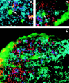
Frozen sections of 12-d embryo thymus with attached mesenchyme. (a) CD45+ stem cells (red) migrating into the epithelial thymus labeled with antibodies to cytokeratin (green). A stem cell can be seen at the mesenchymal–epithelial border (arrow). Fibronectin (blue) is mainly seen over the mesenchymal cells that surround the epithelium, although strong staining is also seen over the endothelial cells of a blood vessel in the upper left corner of the photomicrograph (original magnification: ×400). (b) Staining with the ERTR7 fibroblast marker (red) over mesenchymal cells and capsular material (arrow) surrounding the thymic epithelium (green). Again, fibronectin staining (blue) is mainly seen over mesenchymal cells (original magnification: ×400).
Figure 2
Figure 2
Photograph of lobes cultured with or without mesenchyme. (a) Size comparison between a 12-d thymus lobe (left) with a 15-d lobe (right). The epithelial boundaries of the 12-d lobe (arrows), the attached mesenchyme (M), and a parathyroid gland (P) are indicated. (b) 12-d thymus lobes cultured for 10 d with mesenchyme (top two lobes) or without mesenchyme (bottom three lobes). The scale shows that b is at a lower magnification than a. The parathyroid gland (P) has been left attached to some lobes. There is an obvious size difference between 12-d lobes cultured with mesenchyme and those cultured without.
Figure 3
Figure 3
Flow cytometry of cells in lobes cultured with or without mesenchyme. (a) The proportions of cells expressing CD4 and CD8 in 12-d lobes that have been organ cultured (OC) for 10 d with mesenchyme. Note that the percentages of α/β+ cells in each were as follows: 8.7% (CD4CD8), 31.4% (CD4+CD8+), 31.5% (CD4+CD8), and 12.2% (CD4CD8+). (b) The relatively few CD4+CD8+ cells generated in 12-d lobes cultured without mesenchyme (these cultures yield only 7.7% of the total yield of cells found in cultures with mesenchyme). (c) The CD4 and CD8 phenotypes of cells generated from cultures of 15-d thymus lobes. (d) The development of γ/δ+ cells in cultures of 12-d thymus with mesenchyme. (e) NK1.1+ cells are generated in thymic cultures with mesenchyme. A small proportion of the NK1.1+ cells are also α/β+.
Figure 4
Figure 4
Immunocytochem- istry showing the presence of ERTR7+ mesenchymal cells within the developing and adult thymus. (a) Cells from trypsinized 12-d thymic lobes (epithelium surrounded by mesenchyme). The cells were allowed to attach to wells, and after 48 h were stained for surface fibronectin (green) and, after fixation, for cytokeratin (blue). The image shows cells in phase–contrast with epithelial cells (arrows) and numerous mesenchymal cells. The matrix is associated with the mesenchymal cells. (b and c) Frozen sections of 14-d (not cultured) and 12-d thymus, cultured for 48 h, respectively. ERTR7+ mesenchymal cells (red), some of which are indicated by arrows, can be seen within the thymic epithelium (green). (d) ERTR7+ cells (red) in the cortex of the adult thymus. Some of these cells are present in septae, one of which can be seen on the right of the photomicrograph, whereas others (one marked by an arrow) are present as single cells. Cytokeratin-positive epithelial cells are green and the ECM is blue. Original magnification: (a–d) ×400. (e and f) A single ERTR7+ fibroblast in the adult thymus at a higher magnification (original magnification: ×1,000). In e, the ERTR7 product (red) is indicated by an arrow, and there is a background of cytokeratin-positive epithelial cells (green). The ECM (blue) is associated with the fibroblast. f is a phase contrast view of the same cell (arrow) showing the intimate association with thymocytes.
Figure 5
Figure 5
CFSE-labeled mesenchymal cells migrate into the thymic epithelium of cultured 12-d lobes. (a) CFSE-labeled mesenchymal cells (green) inside the cytokeratin-positive epithelium (blue) of a cultured 12-d lobe. The ERTR7 product is red (arrow). (b and c) CFSE-labeled cells (green, arrow) are distinct from CD45+ lymphoid cells (red). The cytokeratin-positive epithelium is blue. Original magnifications: (a and b) ×1,000; (c) ×800).

References

    1. Cordier A.C., Haumont S.M. Development of thymus, parathyroids and ultimo-branchial bodies in NMRI and nude mice. Am. J. Anat. 1980;157:227–263. - PubMed
    1. Moore M.A.S., Owen J.J.T. Experimental studies on the development of the thymus. J. Exp. Med. 1967;126:715–726. - PMC - PubMed
    1. Le Douarin N.M., Jotereau F.V. Tracing of cells of the avian thymus through embryonic life in interspecific chimeras. J. Exp. Med. 1975;142:17–40. - PMC - PubMed
    1. Anderson G., Moore N.C., Owen J.J.T., Jenkinson E.J. Cellular interactions in thymocyte development. Annu. Rev. Immunol. 1996;14:73–99. - PubMed
    1. Le Lièvre C.S., Le Douarin N.M. Mesenchymal derivatives of the neural crestanalysis of chimeric quail and chick embryos. J. Embryol. Exp. Morphol. 1975;34:125–154. - PubMed

Publication types

MeSH terms