. 2000 Mar 20;191(6):1057-62.
doi: 10.1084/jem.191.6.1057.

Invariant chain controls H2-M proteolysis in mouse splenocytes and dendritic cells

Affiliations

Invariant chain controls H2-M proteolysis in mouse splenocytes and dendritic cells

P Pierre et al. J Exp Med. .

Abstract

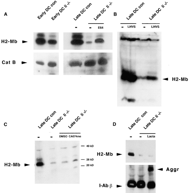
The association of invariant (Ii) chain with major histocompatibility complex (MHC) class II dimers is required for proper antigen presentation to T cells by antigen-presenting cells. Mice lacking Ii chain have severe abnormalities in class II transport, T cell selection, and B cell maturation. We demonstrate here that H2-M, which is required for efficient class II antigenic peptide loading, is unexpectedly downregulated in splenocytes and mature dendritic cells (DCs) from Ii(-/-) mice. Downregulation reflects an increased rate of degradation in Ii(-/-) cells. Degradation apparently occurs within lysosomes, as it is prevented by cysteine protease inhibitors such as E64, but not by the proteasome inhibitor lactacystin. Thus, Ii chain may act as a lysosomal protease inhibitor in B cells and DCs, with its deletion contributing indirectly to the loss of H2-M.


Figures

Figure 2
Figure 2
H2-M is not affected in Ii−/− immature DCs. (A) Immunofluorescence microscopy of bone marrow–derived purified clusters of immature DCs stained for H2-Mb (green) and lamp-2 (red). Control cells (left) and Ii−/− cells (right) do not display any difference for H2-M levels. (B) Confocal immunofluorescence microscopy of immature Langerhans cells from control (left) and Ii−/− mice (right) stained for H2-M (red). No difference in H2-M staining intensity could be detected between these immature Langerhans cells.
Figure 1
Figure 1
H2-M is downregulated in Ii−/− mouse splenocytes. (A) Splenocytes were isolated from control (C57BL/6, Con) or Ii−/− mice. After separation by SDS-PAGE, samples were immunoblotted and H2-Mb was detected using the rabbit polyclonal antibody K553. A severe depletion of H2-M (95%) can be observed in the Ii-deleted mice. (B) Splenocyte extracts from control (C57BL/6), Ii−/−, Ii/MHC class II (CII)−/−, or p31lo mice were immunoblotted for H2-Mb with an anticytoplasmic tail or K553. H2-Mb1 (right) appears as a doublet due its single N-glycosylation detected by the cytoplasmic-tail antibody used here instead of K553, which does not detect this form (left). In Ii chain–deleted mice and double knockout mice, the H2-M level was strongly reduced compared with the control mice (left). However, in p31lo mice (right), H2-M levels were found to be intermediate between control and knockout mice, suggesting a direct correlation between Ii chain levels and H2-M disappearance. (C) To determine if H2-M downregulation was occurring at the transcriptional level, RT-PCR was performed on control, Ii−/−, p31lo, and H2-Ma−/− mice. All levels of transcription for I-Ab α, H2-Mα, and H2-Mβ are found to be identical except in the control mice lacking H2-Mα.
Figure 3
Figure 3
H2-M is downregulated in Ii−/− but not class II−/− mature DCs. Mature LPS-treated control DCs (left) were stained for class II (top, green) and lgp-B/lamp-2 (bottom, red) to demonstrate the maturity of these cells. Control and deleted mature DCs were stained for H2-Mb (top, green) and lgp-B/lamp-2 (bottom, red). In mature DCs, lysosomes converge in the cell center and appear as a unique concentrated H2-M/lamp-2–positive organelle. H2-M is severely downregulated in Ii−/− DCs compared with the control or MHC class II (CII)−/− cells. Lgp-B/lamp-2 levels (right, red) are found to be unchanged in all of the mice strains. H2-Mb downregulation is linked to the absence of Ii and to the maturation of DCs but not to the absence of MHC class II. Some contaminating granulocytes are found to be negative for H2-M in control cells and class II−/−. Bars, 100 μM.
Figure 4
Figure 4
Quantitation of H2-M half-life. (A) Immunoprecipitation of S35-pulsed and chased H2-M with the mAb 2C3A was performed. H2-Mb was found to be degraded almost completely after 24 h in Ii−/− DCs in contrast to the control (C) DCs. (B) Quantitation of H2-Mb immunoprecipitated with 2C3A (n = 2) from control (Con) or Ii−/− late DCs treated or not with LHVS (1 μM) during the chase. A clear difference can be observed after 24 h of chase between the control and Ii−/− DCs. Interestingly, LHVS treatment during the chase prevents H2-Mb degradation, suggesting a cysteine protease involvement. (C and D) In control DCs, the p41 alternatively spliced form of Ii chain is preferentially coimmunoprecipitated with H2-M (2C3A) after 30 min of radioactive labeling, whereas p31 is the main Ii chain isoform immunoprecipitated using the IN1 mAb.
Figure 5
Figure 5
Cysteine proteases inhibition prevents H2-M degradation in mature DCs. (A) Early and late (LPS-treated) DCs from control (con) or Ii−/− mice were purified and treated with protease inhibitor before immunoblot for H2-Mb and cathepsin B. H2-Mb was mildly downregulated in Ii−/− early DCs and virtually absent from Ii−/− late DCs. Incubation of these cells with the cysteine protease inhibitor E64 for 24 h allowed for an efficient recovery of H2-M. Little variation in cathepsin B expression was observed between control and Ii-deleted cells (bottom) or during maturation. This observation confirms that H2-M is actively degraded by cysteine proteases in the endocytic pathway of late DCs. (B) LHVS at 1 μM but not at 2 nM (not shown) had the same effect as E64, suggesting that lysosomal cysteine proteases but not cathepsin S are degrading H2-M. (C) Ii−/− DCs were treated for 18 h with 5 μM CA074me, resulting in partial H2-M rescue. The specificity of CA074 for cathepsin B suggests a role of this protease in H2-M degradation. (D) Ii−/− DCs were treated for 18 h with 50 μM lactacystin, with no effect on H2-M disappearance. MHC class II (I-Ab) molecules were detected to demonstrate accumulation of class II aggregates (Aggr) in the lactacystin (Lacta)-treated cells.

References

    1. Cresswell P. Assembly, transport, and function of MHC class II molecules. Annu. Rev. Immunol. 1994;12:259–293. - PubMed
    1. Wolf P.R., Ploegh H.L. How MHC class II molecules acquire peptide cargobiosynthesis and trafficking through the endocytic pathway. Annu. Rev. Cell Biol. 1995;11:267–306. - PubMed
    1. Cresswell P. Invariant chain structure and MHC class II function. Cell. 1996;84:505–507. - PubMed
    1. Roche P.A. HLA-DMan in vivo facilitator of MHC class II peptide loading. Immunity. 1995;3:259–262. - PubMed
    1. Kropshofer H., Hammerling G.J., Vogt A.B. How HLA-DM edits the MHC class II peptide repertoiresurvival of the fittest? Immunol. Today. 1997;18:77–82. - PubMed

Publication types

MeSH terms