. 2000 Apr;74(8):3441-8.
doi: 10.1128/jvi.74.8.3441-3448.2000.

Synaptic pathology in Borna disease virus persistent infection

Affiliations

Synaptic pathology in Borna disease virus persistent infection

D Gonzalez-Dunia et al. J Virol. 2000 Apr.

Abstract

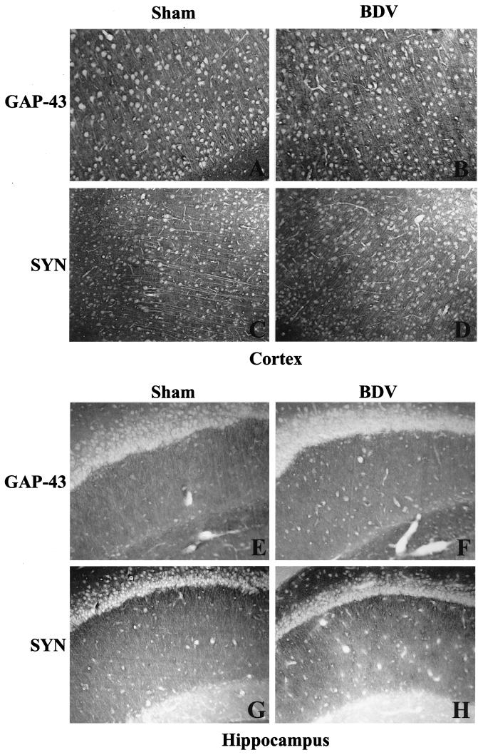
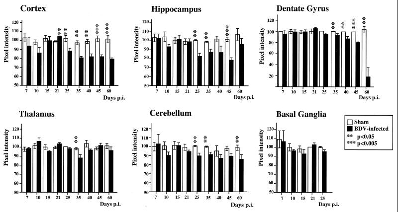
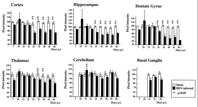
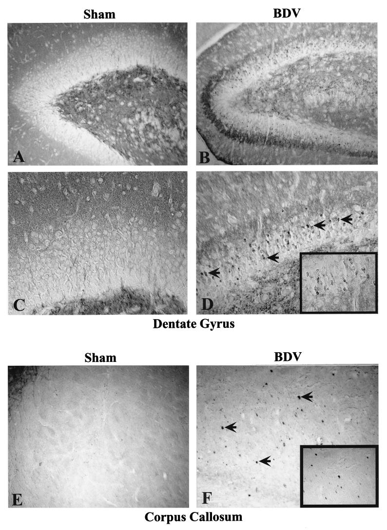
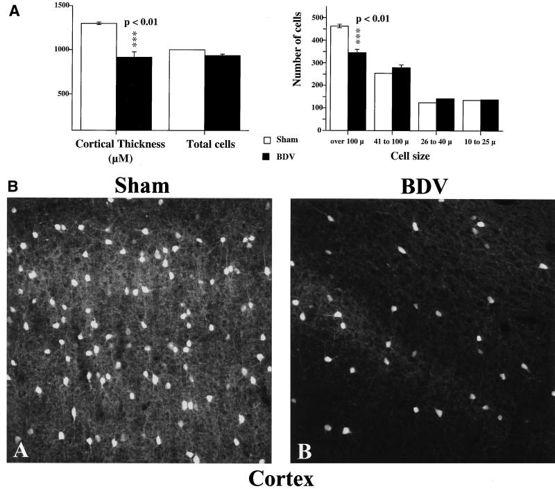
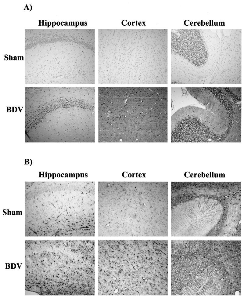
Borna disease virus (BDV) infection of newborn rats leads to a persistent infection of the brain, which is associated with behavioral and neuroanatonomical abnormalities. These disorders occur in the absence of lymphoid cell infiltrates, and BDV-induced cell damage is restricted to defined brain areas. To investigate if damage to synaptic structures anteceded neuronal loss in BDV neonatally infected rats, we analyzed at different times postinfection the expression levels of growth-associated protein 43 and synaptophysin, two molecules involved in neuroplasticity processes. We found that BDV induced a progressive and marked decrease in the expression of these synaptic markers, which was followed by a significant loss of cortical neurons. Our findings suggest that BDV persistent infection interferes with neuroplasticity processes in specific cell populations. This, in turn, could affect the proper supply of growth factors and other molecules required for survival of selective neuronal populations within the cortex and limbic system structures.


Figures

FIG. 1
FIG. 1
Distribution of GAP-43 and SYN IR in sham- and neonatally BDV-infected rats, 40 days p.i. Cortex (A to D) and hippocampus (E to H) from control and infected rats exhibit the characteristic granular immunolabeling of the neuropil but not the cell bodies. Magnification, ×100.
FIG. 2
FIG. 2
Quantitative analysis of GAP-43 IR in the cortex, hippocampus, DG, thalamus, cerebellum, and basal ganglia from control and BDV-infected rats. Values of pixel intensities (percentage of median) are mean ± SEM, each bar representing the result of at least eight independent measurements. Double (P < 0.05) and triple (P < 0.005) asterisks indicate the level of statistical significance (determined by the Mann-Whitney U test). Loss of GAP-43 IR in the DG of neonatally infected rats followed the kinetics of DG granule cell degeneration observed in these animals. Compared to age-matched controls, GAP-43 levels are significantly reduced in the cortex and hippocampus starting at about day 25 p.i. No significant differences are observed elsewhere.
FIG. 3
FIG. 3
Quantitative analysis of SYN IR in the cortex, hippocampus, DG, thalamus, cerebellum, and basal ganglia from control and BDV-infected rats. Values of pixel intensities (percentage of median) are mean ± SEM, each bar representing the result of at least eight independent measurements. Double asterisks (P < 0.05) indicate the level of statistical significance (determined by the Mann-Whitney U test). Loss of SYN IR in the DG of neonatally infected rats precedes and follows the kinetics of DG granule cell degeneration observed in these animals. Compared to age-matched controls, SYN levels are significantly reduced in the cortex, hippocampus, and thalamus.
FIG. 4
FIG. 4
Abnormal SYN immunostaining in BDV-infected rat brain. Note the clustering of SYN IR within the inner molecular layer of the DG at 25 days p.i. (compare panels A and B). SYN staining is also found in the cell bodies of dentate granule cells (arrowed cells in panel D, shown at a larger magnification in the boxed area). By day 45 p.i., the corpus callosum, which is usually negative for SYN staining in control rats (panel E), exhibits a patched IR in infected animals (arrows in panel F and boxed area). The immunostaining accumulates in axonal blobs, indicating an abnormal axoplasmic flow and a poor transport of SYN to the synaptic terminals. Magnification, ×150.
FIG. 5
FIG. 5
Cell loss in the cortex of neonatally BDV-infected animals, 45 days p.i. (A) Morphometric analysis. Results are expressed as mean ± SEM. Averages are based on results from five each sham-inoculated and rats with BDV PTI (two sections per animal). The level of statistical significance (determined by Mann-Whitney U test) is indicated by triple asterisk (P < 0.01). BDV-infected rats display significant cortical shrinkage (about 30%) and loss of cells with a diameter of >100 μm compared to noninfected age-matched controls. (B) Immunohistochemical staining for parvalbumin in the cortex of sham- and BDV-infected rats, 45 days p.i. Parvalbumin, a calcium-binding protein, labels GABA-ergic neurons in the cortex. Similar cortical areas are shown for each animal. Note the significant decrease in numbers of stained cells and processes in the infected animal. Magnification, ×250.
FIG. 6
FIG. 6
Detection of BDV antigen and GFAP expression in control and infected rat brains, 35 days p.i. (A) Expression of BDV nucleoprotein. Sections were immunostained with an antibody specific for BDV nucleoprotein. There is a strong nuclear staining in abundant pyramidal cells of the hippocampus and neocortex, as well as in Purkinje and granule cells of the cerebellum. Diffuse staining of the neuropil is also observed. There was no staining in sham-inoculated animals. Magnification, ×80. (B) Analysis of GFAP expression in similar fields. Neonatally infected rats display a significant increase in the number of GFAP-positive astroglial cells in the hippocampus (molecular layer and dentate gyrus), cortex, and cerebellum. Consistent with this activation, astrocytes are significantly hypertrophied in infected brains. Magnification, ×80.

References

    1. Bautista J R, Rubin S A, Moran T H, Schwartz G J, Carbone K M. Developmental injury to the cerebellum following perinatal Borna disease virus infection. Dev Brain Res. 1995;90:45–53. - PubMed
    1. Bautista J R, Schwartz G J, de la Torre J C, Moran T H, Carbone K M. Early and persistent abnormalities in rats with neonatally acquired Borna disease virus infection. Brain Res Bull. 1994;34:31–36. - PubMed
    1. Bode L. Human infections with Borna disease virus and potential pathogenic implications. In: Koprowski H, Lipkin I, editors. Borna disease. Berlin, Germany: Springer-Verlag KG; 1995. pp. 103–130. - PubMed
    1. Bode L, Zimmermann W, Ferszt R, Steinbach F, Ludwig H. Borna disease virus genome transcribed and expressed in psychiatric patients. Nat Med. 1995;1:232–236. - PubMed
    1. Brahic M, Ozden S. Simultaneous detection of cellular RNA and proteins. In: Wilkinson R G, editor. In situ hybridization—a practical approach. Oxford, England: IRL Press; 1992. pp. 85–104.

Publication types

LinkOut - more resources