. 2000 Apr;74(8):3470-7.
doi: 10.1128/jvi.74.8.3470-3477.2000.

NP and L proteins of lymphocytic choriomeningitis virus (LCMV) are sufficient for efficient transcription and replication of LCMV genomic RNA analogs

Affiliations

NP and L proteins of lymphocytic choriomeningitis virus (LCMV) are sufficient for efficient transcription and replication of LCMV genomic RNA analogs

K J Lee et al. J Virol. 2000 Apr.

Abstract

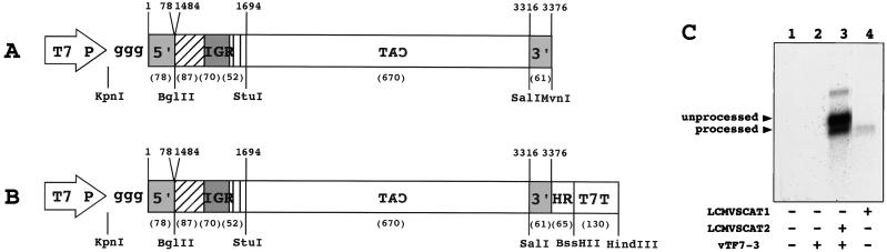
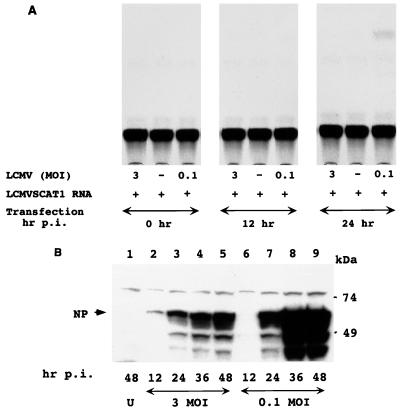
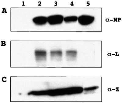
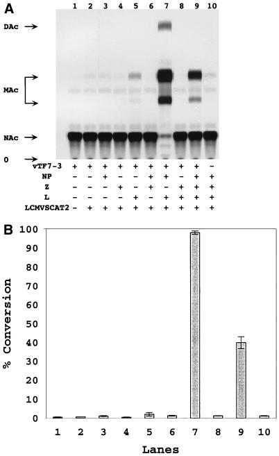
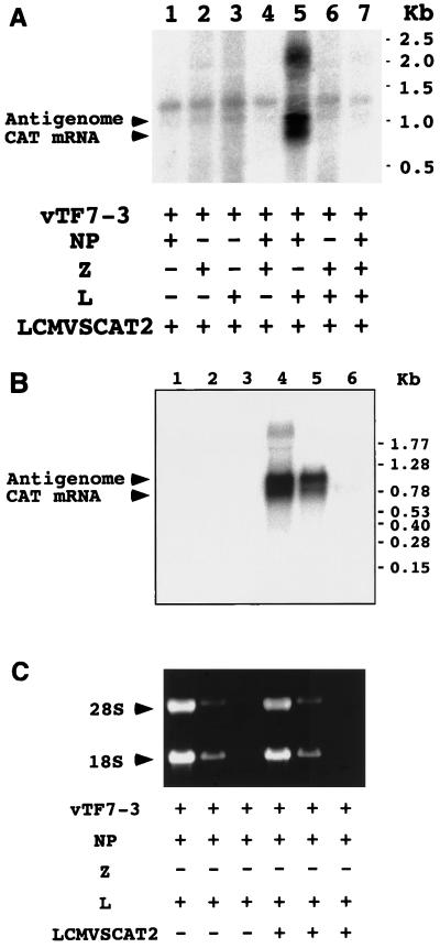
The genome of lymphocytic choriomeningitis virus (LCMV) consists of two negative-sense single-stranded RNA segments, designated L and S. Both segments contain two viral genes in an ambisense coding strategy, with the genes being separated by an intergenic region (IGR). We have developed a reverse genetic system that allows the investigation of cis-acting signals and trans-acting factors involved in transcription and replication of LCMV. To this end, we constructed an LCMV S minigenome consisting of a negative-sense copy of the chloramphenicol acetyltransferase (CAT) reporter gene flanked upstream by the S 5' untranslated region (UTR) and IGR and downstream by the S 3' UTR. CAT expression was detected in LCMV-infected cells transfected with the minigenome RNA. Intracellular coexpression of the LCMV minigenome and LCMV L and NP proteins supplied from cotransfected plasmids driven by the T7 RNA polymerase provided by the recombinant vaccinia virus vTF7-3 resulted in high levels of CAT activity and synthesis of subgenomic CAT mRNA and antiminigenome RNA species. Thus, L and NP represent the minimal viral trans-acting factors required for efficient RNA synthesis mediated by LCMV polymerase.


Figures

FIG. 1
FIG. 1
Schematic diagrams and characterization of LCMV genomic RNA analogs. (A) Plasmid pLCMVSCAT1 was constructed by combining, in the order indicated in the schematic, the following elements: the minimum T7 RNA polymerase promoter (T7 P) followed by three extra G's, the 5′ UTR of the LCMV S RNA (nt 1 to 78), the IGR of the LCMV S RNA (nt 1484 to 1694), a DNA encoding the full-length CAT ORF in antisense orientation with respect to the T7 promoter, and the 3′ UTR of the LCMV S RNA (nt 3316 to 3376). (B) Plasmid pLCMVSCAT2 was made by inserting an LCMV-specific 3′ HR followed by T7T into pLCMVSCAT1. Viral sequences were derived from LCMV Arm 5. Nucleotide numbers correspond to those of the S RNA. Numbers in parentheses indicate lengths of cDNA fragments. Restriction enzymes used for cloning are indicated at the bottom. Hatched and striped boxes represent the 3′ ends of the GP and NP, respectively. (C) Northern blot analysis of in vitro and intracellularly synthesized LCMV RNA minigenomes. BHK-21 cells were infected with vTF7-3 at an MOI of 3 PFU/cell and subsequently transfected with 2 μg of pLCMVSCAT2. Total cellular RNA was prepared by using TRI reagent at 24 h after infection with vTF7-3. RNA (5 μg) was analyzed by Northern blotting using a CAT sense riboprobe. LCMVSCAT1 RNA was prepared in vitro transcription with T7 RNA polymerase of MvnI-digested template pLCMVSCAT1 DNA. Unprocessed and ribozyme-processed RNA species are indicated by arrowheads at the left.
FIG. 2
FIG. 2
Rescue of CAT activity by LCMV helper virus correlates with expression of viral NP. (A) Helper virus-dependent rescue of CAT expression. BHK-21 cells were infected with LCMV at an MOI of either 0.1 or 3 and then transfected with 5 μg of in vitro-transcribed LCMVSCAT1 RNA at 0, 12, and 24 p.i. Cells were harvested at 48 h p.i., and extracts for CAT assay were prepared. CAT assay was performed as described in Materials and Methods. Shown is a representative result of four independent experiments. (B) Kinetics of NP expression in LCMV-infected BHK-21 cells. BHK-21 cells were infected with LCMV at an MOI of either 0.1 or 3. Cells were harvested at 12, 24, 36, and 48 h p.i. Uninfected BHK-21 control cells were harvested at 48 h p.i. Similar amounts of whole cell extracts from each sample were analyzed by Western blotting with a monoclonal antibody to NP. Immunoreactivity was detected by chemiluminescence. U, uninfected cells. Numbers at the bottom correspond to time of harvest. Molecular weight standards are indicated on the right. The position of NP is indicated by an arrow on the left. Consistent with previous reports, additional bands corresponding to specific NP fragments could be seen in extracts from LCMV-infected cells.
FIG. 3
FIG. 3
Plasmid-mediated expression of LCMV trans-acting factors. BHK-21 cells (4 × 10⁵) were infected with vTF7-3 at an MOI of 3 and then transfected with pCITE-NP, pUCIRES-Z, and pGEM-L. Transfected cells were harvested at 24 h p.i. BHK-21 cells (2 × 10⁵) were infected with LCMV at an MOI of 0.1 PFU/cell and harvested at 48 h p.i. Whole cell extracts were separated by 10, 16, and 6% SDS-PAGE for NP, Z, and L (top, middle, and bottom, respectively). Monoclonal antibody to NP and polyclonal antibody to Z and L were used for detection of LCMV proteins by Western blotting using chemiluminescence. Lanes: 1, cells infected with vTF7-3, 2, cells infected with vTF7-3 and transfected with 0.4 μg of either pCITE-NP, pUCIRES-Z, or pGEM-L; 3, BHK-21 cells infected with vTF7-3 and transfected with 1.2 μg of either pCITE-NP, pUCIRES-Z, or pGEM-L; 4, BHK-21 cells infected with vTF7-3 and cotransfected with 0.4 μg of each of plasmids pCITE-NP, pUCIRES-Z, and pGEM-L; 5, cells infected with LCMV at an MOI of 0.1. Antibodies used are indicated at the right.
FIG. 4
FIG. 4
Expression of LCMV minigenome in cells infected with vTF7-3 and transfected with pLCMVSCAT2 and LCMV NP, Z, and L protein expression plasmids. (A) Analysis of CAT activity. BHK-21 cells (4 × 10⁵) were infected with vTF7-3 at an MOI of 3 PFU/cell and then transfected with various combination of plasmids pLCMVSCAT2, pCITE-NP, pUCIRES-Z, and pGEM-L as indicated in Materials and Methods. Cells were harvested at 24 h p.i. and analyzed for CAT activity by TLC as described in Materials and Methods. + and − indicate presence and the absence of plasmid or vTF7-3. NAc, MAc, and DAc indicate the positions of nonacetylated, monoacetylated, and diacetylated forms, respectively, of chloramphenicol in the TLC; O indicates the origin of the TLC. (B) CAT activities were quantified as described in Materials and Methods and presented as percent conversion from nonacetylated to acetylated forms of chloramphenicol. Values correspond to average ± standard deviation of three independent experiments.
FIG. 5
FIG. 5
Analysis of RNA synthesized intracellularly by LCMV polymerase components. (A) Northern blot analysis of total cellular RNA. BHK-21 cells were infected with vTF7-3 at an MOI of 3 and then transfected with pLCMVSCAT2 and LCMV NP, Z, and L protein expression plasmids as for Fig. 4. Cells were harvested at 24 h p.i., and total RNA was isolated. RNA (5 μg) was analyzed by Northern blotting using an antisense CAT riboprobe. + and − indicate presence and absence of plasmid or vTF7-3. CAT mRNA and antigenome RNA are indicated by arrowheads on the left. (B) Oligo(dT) chromatography analysis. RNA isolated from BHK-21 cells infected with vTF7-3 transfected with the indicated plasmids was fractionated by oligo(dT) chromatography. Total (lanes 1 and 4), poly(A) (lanes 2 and 5), and poly(A)+ (lanes 3 and 6) RNAs corresponding to equivalent amounts of cell numbers were analyzed by Northern blot hybridization using an antisense CAT riboprobe. (C) Ethidium bromide staining of the formaldehyde agarose gel prior transfer of the RNA samples to the membrane.

References

    1. Atreya P L, Peeples M E, Collins P L. The NS1 protein of human respiratory syncytial virus is a potent inhibitor of minigenome transcription and RNA replication. J Virol. 1998;72:1452–1461. - PMC - PubMed
    1. Auperin D D, Compans R W, Bishop D H. Nucleotide sequence conservation at the 3′ termini of the virion RNA species of New World and Old World arenaviruses. Virology. 1982;121:200–203. - PubMed
    1. Auperin D D, Romanowski V, Galinski M, Bishop D H. Sequencing studies of pichinde arenavirus S RNA indicate a novel coding strategy, an ambisense viral S RNA. J Virol. 1984;52:897–904. - PMC - PubMed
    1. Auperin D D, Sasso D R, McCormick J B. Nucleotide sequence of the glycoprotein gene and intergenic region of the Lassa virus S genome RNA. Virology. 1986;154:155–167. - PubMed
    1. Borden K L, Campbell Dwyer E J, Salvato M S. An arenavirus RING (zinc-binding) protein binds the oncoprotein promyelocyte leukemia protein (PML) and relocates PML nuclear bodies to the cytoplasm. J Virol. 1998;72:758–766. - PMC - PubMed

Publication types

MeSH terms

LinkOut - more resources