

doi: 10.1128/jvi.74.8.3709-3714.2000.
A novel subgenomic murine leukemia virus RNA transcript results from alternative splicing
Affiliations
- PMID: 10729146
- PMCID: PMC111880
- DOI: 10.1128/jvi.74.8.3709-3714.2000
Item in Clipboard
A novel subgenomic murine leukemia virus RNA transcript results from alternative splicing
J Virol.
2000 Apr.
Abstract
Here we show the existence of a novel subgenomic 4.4-kb RNA in cells infected with the prototypic replication-competent Friend or Moloney murine leukemia viruses (MuLV). This RNA derives by splicing from an alternative donor site (SD') within the capsid-coding region to the canonical envelope splice acceptor site. The position and the sequence of SD' was highly conserved among mammalian type C and D oncoviruses. Point mutations used to inactivate SD' without changing the capsid-coding ability affected viral RNA splicing and reduced viral replication in infected cells.
Figures
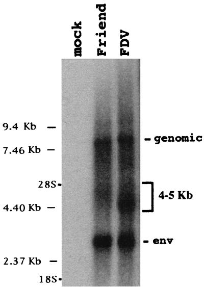
Viral RNA species expressed in MuLV-infected cells. Cellular RNA from mock-infected cells or cells acutely infected with wt Friend MuLV or the FDV mutant (see Fig. 2) were analyzed by Northern blot hybridization with a radiolabeled probe complementary to the 3′ MuLV region (nt 5638 to 8327). This probe reveals the viral genomic RNA (8.3 kb), the single-spliced env RNA (3 kb), and a third RNA species (4 to 5 kb). Positions of RNA molecular size markers and 28S and 18S ribosomal RNAs are indicated.
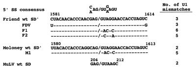
Sequences of Friend and Moloney MuLV mutants in the SD′ region. Sequences of Friend (FDV, F1, and F2) and Moloney (M1) viruses containing changes in the SD′ region were aligned with the 5′ splice donor site (5′ SS) consensus sequence of U1 snRNA. The cleavage site is indicated by a slash mark. The nucleotide changes maintain the coding potential of the parental strains and are indicated for each mutant. For comparison, the sequence of the conserved canonical MuLV 5′ splice donor site (SD) is shown. The number of potential mismatches between each splice donor site and the U1 snRNA is on the right.
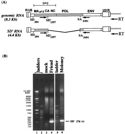
Amplification of a new alternatively spliced MuLV transcript by RT-PCR. (A) Schematic structures of the unspliced genomic and alternatively spliced (SD′) RNA, including the canonical (SD), acceptor (SA), and alternative (SD′) splice sites. Nucleotides are numbered starting from the first nucleotide of R according to the Friend MuLV sequence. Also noted are the gag gene components, including the matrix (MA), capsid (CA), and nucleocapsid (NC). Arrows refer to the approximate positions of primers used for RT with the oligo(dT) primer and for PCR amplification. (B) RT-PCR was conducted on total RNA samples extracted from mock-infected cells (lane 3) or cells infected with either Friend (lane 4) or Moloney virus (lane 6). Reverse transcriptions were performed with oligo(dT), and PCRs were performed with oligonucleotides s1450 and a5620 as described in the Materials and Methods. The alternatively spliced SD′ RNA yielded an amplified product of 276 bp. After 30 cycles, amplified samples were loaded onto an agarose gel and stained with ethidium bromide. Lanes 1 and 2, 1-kb and 100-bp ladders, respectively.
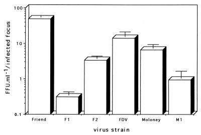
Replication of wt and mutant viruses. FIA was used to quantitate infectious virions present in the supernatants harvested from cell cultures with the same number of wt or mutant MuLV-infected foci. Infectivities were performed at least three times for each virus in parallel and each test was performed in triplicate. Bars, the standard error of the mean of each series.
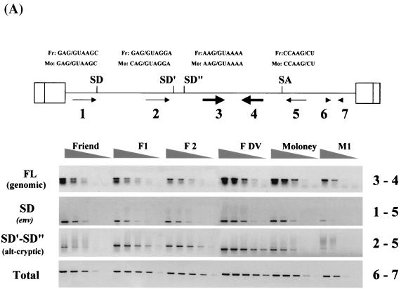
Effect of SD′ mutations on MuLV splicing. The different RNA species were detected by semiquantitative PCRs (20 to 24 cycles) performed on threefold dilutions of each sample. The amplified products were run onto agarose gels containing ethidium bromide. (A) Schematic MuLV genome map indicating the splice donor (SD, SD′, and SD") and acceptor (SA) sites. The corresponding sequences in Friend (Fr) and Moloney (Mo) are shown in the upper panel. Approximate positions of the primers used to amplify the different RNA species are identified by the numbered arrows. Each viral RNA species detected is indicated on the left side of the gel, with the corresponding oligonucleotide pairs identified by numbers on the right. Note that the main products amplified from the F1, F2, and M1 mutants correspond to usage of the cryptic SD" and exhibit slower migration relative to products arising from usage of the alternative SD′ in the wt and FDV. (B) Quantification of the different RNA species was determined by a FluorImager. For each virus, at least two RNA preparations were isolated and subjected to three to six semiquantitative RT-PCR analyses. For each RNA species, the RNA levels are expressed as the ratio of the value obtained after amplification with the specific oligonucleotide pair divided by the total RNA value obtained by amplification with primers 6 and 7. Final RNA levels are represented after normalization to levels of the corresponding wt strain (± standard error of the mean). Note that both wt Friend and Moloney, as well as the FDV mutant, produced alternatively SD′ spliced RNA, while F1, F2, and M1 mutants displayed a spliced RNA resulting from the activation of the SD" cryptic splice site.
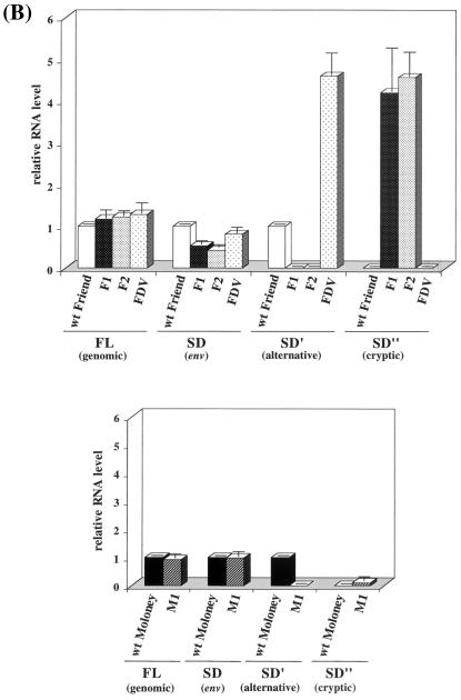
Effect of SD′ mutations on MuLV splicing. The different RNA species were detected by semiquantitative PCRs (20 to 24 cycles) performed on threefold dilutions of each sample. The amplified products were run onto agarose gels containing ethidium bromide. (A) Schematic MuLV genome map indicating the splice donor (SD, SD′, and SD") and acceptor (SA) sites. The corresponding sequences in Friend (Fr) and Moloney (Mo) are shown in the upper panel. Approximate positions of the primers used to amplify the different RNA species are identified by the numbered arrows. Each viral RNA species detected is indicated on the left side of the gel, with the corresponding oligonucleotide pairs identified by numbers on the right. Note that the main products amplified from the F1, F2, and M1 mutants correspond to usage of the cryptic SD" and exhibit slower migration relative to products arising from usage of the alternative SD′ in the wt and FDV. (B) Quantification of the different RNA species was determined by a FluorImager. For each virus, at least two RNA preparations were isolated and subjected to three to six semiquantitative RT-PCR analyses. For each RNA species, the RNA levels are expressed as the ratio of the value obtained after amplification with the specific oligonucleotide pair divided by the total RNA value obtained by amplification with primers 6 and 7. Final RNA levels are represented after normalization to levels of the corresponding wt strain (± standard error of the mean). Note that both wt Friend and Moloney, as well as the FDV mutant, produced alternatively SD′ spliced RNA, while F1, F2, and M1 mutants displayed a spliced RNA resulting from the activation of the SD" cryptic splice site.
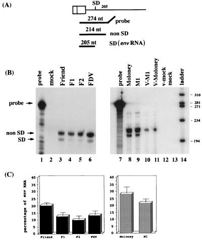
Quantification by RNase protection of the canonical env transcript level in MuLV-infected cells. (A) Friend and Moloney transcripts were detected by RNase protection with the uniformly labeled antisense SPFLV or SPMLV probes, respectively (see Materials and Methods), which overlap the canonical SD site. Hybridization of the 274-nt probe to viral RNA species that are not spliced at the canonical SD site (full length, SD′, and SD" RNA) yields a protected 214-nt fragment, while canonically spliced env RNA yields a protected 205-nt fragment. (B) RNase protection assays were carried out with 15 μg of total cellular RNA (lanes 2 to 6, 8, 9, and 13) and RNA extracted from 10% total virus pelleted medium from infected or mock-infected cell monolayers (lanes 10 to 12). The positions of the probe and the protected fragments corresponding to canonically spliced env RNA (SD) as well as the noncanonically spliced RNAs (non SD) are indicated by arrows. The size markers (lane 14) consist of end-labeled φX174 HaeIII DNA fragments. (C) Quantification of RNase protection assays. For each series, the percentage of env-protected fragments versus the total of protected signals is represented (± standard of the mean). Each value corresponds to the average of at least three measurements performed on different RNA preparations.
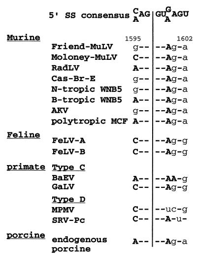
Conservation of the SD′ site. Sequence alignment of a putative SD′ site in the capsid-coding region of a series of replication-competent mammalian types C and D retroviruses. The 5′ splice donor site consensus sequence (5′ SS) is shown on top, with the potential splicing cleavage site indicated by a vertical line. All sequences were located approximately 100 nt upstream of the capsid major homology region. Numbering is according to the Friend-MuLV 57 sequence. Lower-case letters indicate mismatches between the 5′SS consensus and the viral sequence. Abbreviations and strains correspond to the following retroviruses. (i) MuLVs: Friend-MuLV, strain 57; Moloney-MuLV, strain 8.2; RadLV, radiation leukemia virus; Cas-Br-E, Lake Casitas brain E neurotropic virus; WNB5, the N- and B-tropic clones of the WN1802 isolate; and AKV, from clone AKR 623 of endogenous virus from the AKR mouse strain. All of the above are ecotropic MuLVs. MCF, clone MCF1233 of the polytropic mink cell focus-inducing viruses. (ii) Feline leukemia viruses: FeLVA and FeLVB, strains A and B. (iii) Primate simple retroviruses: simian type C retroviruses include BaEV, baboon endogenous virus, and GaLV, gibbon ape leukemia virus. Simian type D retroviruses include MPMV, Mason-Pfizer monkey virus, and SRV-Pc, a baboon simian retrovirus-like isolate. (iv) Porcine endogenous virus: a human-tropic C-type porcine endogenous retrovirus.
References
-
- Barklis E, Mulligan R C, Jaenisch R. Chromosomal position or virus mutation permits retrovirus expression in embryonal carcinoma cells. Cell. 1986;47:391–399. - PubMed
-
- Eiden M, Trainor C D, Reitz M S. Gibbon ape leukaemia virus RNA in leukaemic T-lymphoid cell lines: expression of a novel RNA transcript. J Gen Virol. 1986;67:1455–1460. - PubMed
Publication types
MeSH terms
Substances
LinkOut - more resources
Full Text Sources
Other Literature Sources
