

doi: 10.1128/jvi.74.8.3881-3887.2000.
Induction of a novel cellular homolog of interleukin-10, AK155, by transformation of T lymphocytes with herpesvirus saimiri
Affiliations
- PMID: 10729163
- PMCID: PMC111897
- DOI: 10.1128/jvi.74.8.3881-3887.2000
Item in Clipboard
Induction of a novel cellular homolog of interleukin-10, AK155, by transformation of T lymphocytes with herpesvirus saimiri
J Virol.
2000 Apr.
Abstract
Although herpesvirus saimiri-transformed T lymphocytes retain multiple normal T-cell functions, only a few changes have been described. By subtractive hybridization, we have isolated a novel cellular gene, ak155, a sequence homolog of the interleukin-10 gene. Specifically herpesvirus saimiri-transformed T cells overexpress ak155 and secrete the protein into the supernatant. In other T-cell lines and in native peripheral blood cells, but not in B cells, ak155 is transcribed at low levels. AK155 forms homodimers similarly to interleukin-10. As a lymphokine, AK155 may contribute to the transformed phenotype of human T cells after infection by herpesvirus saimiri.
Figures
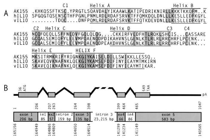
Amino acid sequence alignment and genomic structure of ak155. (A) The amino acid sequences of AK155, human IL-10 (hIL10), and EBV IL-10 (vIL10) were aligned. Identical amino acids are shaded. Cysteine residues C1 to C4 are conserved. Six predicted helical areas (helices A to F) for the three proteins are marked. (B) The genomic exon-intron structure and the ak155 coding region are depicted. The nucleotide positions above the structure refer to ak155 cDNA. The nucleotide positions below the structure correspond to the genomic sequence of the human chromosome 12q15 region.
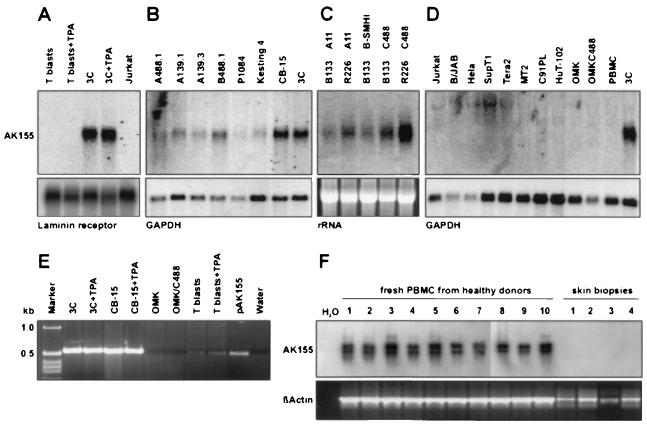
ak155 transcription pattern. The transcription of ak155 was analyzed by Northern blotting (A to D) and RT-PCR (E and F). (A) Strong ak155 transcript bands were demonstrated for the HVS-transformed CD8+ human T-cell line 3C, but not for either nontransformed T cells or Jurkat cells. Phorbol ester stimulation (TPA; 2 ng/ml for 6 h) did not affect ak155 transcription. Laminin receptor transcripts are shown as a control. (B) A series of additional HVS-transformed T-cell lines also transcribed ak155: CB-15, Kesting, and A488.1 (CD4+; transformed by C488), P1084 and B488.1 (CD8+; transformed by C488), and the C139-transformed T-cell lines A139.1 (γδ T-cell receptor) and A139.3 (αβ, CD4+). Glyceraldehyde-3-phosphate dehydrogenase (GAPDH) transcripts are shown as a control. (C) ak155 transcripts were also demonstrated in transformed T cells from New World monkeys (S. oedipus) (T cells from donors B133 and R226 [24]). ak155 expression was not specific for the virus subgroup used for transformation and was similarly detectable in T cells transformed by the virus strains A11, B-SMHI, and C488. rRNA bands are shown as a transfer control. (D) Various other cell types were tested by Northern blotting for ak155 transcripts (Jurkat, SupT1, MT2, C91PL, HuT-102, B/JAB, HeLa, and Tera2). No additional ak155-positive lines were identified. Infection of the permissive epithelial cell line OMK with HVS C488 did not induce ak155 transcription. GAPDH transcripts are shown as a control. (E) By using RT-PCR we confirmed that HVS-transformed human T-cell lines (CB-15 and 3C) transcribed ak155 at a high level (540-bp fragment). Transcripts were also detected in T blasts but at low levels. Additional phorbol ester stimulation did not change the signal intensity. The cDNA plasmid pAK155 served as a positive control. (F) Weak RT-PCR signals for ak155 transcripts were detected from unstimulated fresh peripheral blood mononuclear cells (PBMC) of 10 healthy blood donors by ethidium bromide staining and confirmed by Southern blot hybridization. β-Actin transcripts are shown as a positive control.
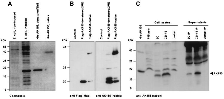
AK155 dimerization and production by HVS-transformed human T cells. (A) Recombinant amino-terminally histidine-tagged AK155 protein was expressed after induction in E. coli. The protein was purified by nickel-nitrilotriacetic acid-agarose chromatography. The denatured recombinant protein was demonstrated as a 19-kDa band in SDS gels. In absence of 2-mercaptoethanol (2ME) and without heat denaturation, the 19-kDa band shifted to the 36-kDa position. A Coomassie-stained SDS gel is shown. (B) Recombinant amino-terminally Flag-tagged AK155 protein was expressed in COS-7 cells after transfection. The recombinant protein was easily detectable by Western blotting either with the anti-Flag monoclonal antibody or with rabbit antiserum. In both cases, the eukaryotically expressed protein efficiently formed dimers when tested under nondenaturing conditions. (C) The endogenous AK155 protein from HVS-transformed human T cells (3C and CB-15) was demonstrated by Western blotting with rabbit antiserum. Protein was detected in lysates of the transformed T-cell lines 3C and CB-15 without previous immunoprecipitation and in their supernatants after immunoprecipitation with rabbit antiserum and protein G-agarose. As a control, bacterially expressed His-AK155 is shown in the first lane. Due to the histidine tag, this protein appears at a slightly larger size than the endogenous protein from 3C and CB-15 cells. AK155 was detectable neither in Jurkat cells nor in their supernatant. The Western blots were developed with chemiluminescence reactions.
References
-
- Biesinger B, Tsygankov A Y, Fickenscher H, Emmrich F, Fleckenstein B, Bolen J B, Bröker B M. The product of the herpesvirus saimiri open reading frame 1 (tip) interacts with T cell-specific kinase p56lck in transformed cells. J Biol Chem. 1995;270:4729–4734. - PubMed
-
- Bröker B M, Tsygankov A Y, Müller-Fleckenstein I, Guse A H, Chitaev N A, Biesinger B, Fleckenstein B, Emmrich F. Immortalization of human T cell clones by herpesvirus saimiri. Signal transduction analysis reveals functional CD3, CD4 and IL-2 receptors. J Immunol. 1993;151:1184–1192. - PubMed
-
- Bröker B M, Fickenscher H. Herpesvirus saimiri strategies for T cell stimulation and transformation. Med Microbiol Immunol. 1999;187:127–136. - PubMed
Publication types
MeSH terms
Substances
Associated data
- Actions
- Actions
LinkOut - more resources
Full Text Sources
Other Literature Sources
Medical
Molecular Biology Databases
Research Materials
