. 2000 Apr;84(4):408-12.
doi: 10.1136/bjo.84.4.408.

Both CD4+ and CD8+ T cells are involved in protection against HSV-1 induced corneal scarring

Affiliations

Both CD4+ and CD8+ T cells are involved in protection against HSV-1 induced corneal scarring

H Ghiasi et al. Br J Ophthalmol. 2000 Apr.

Abstract

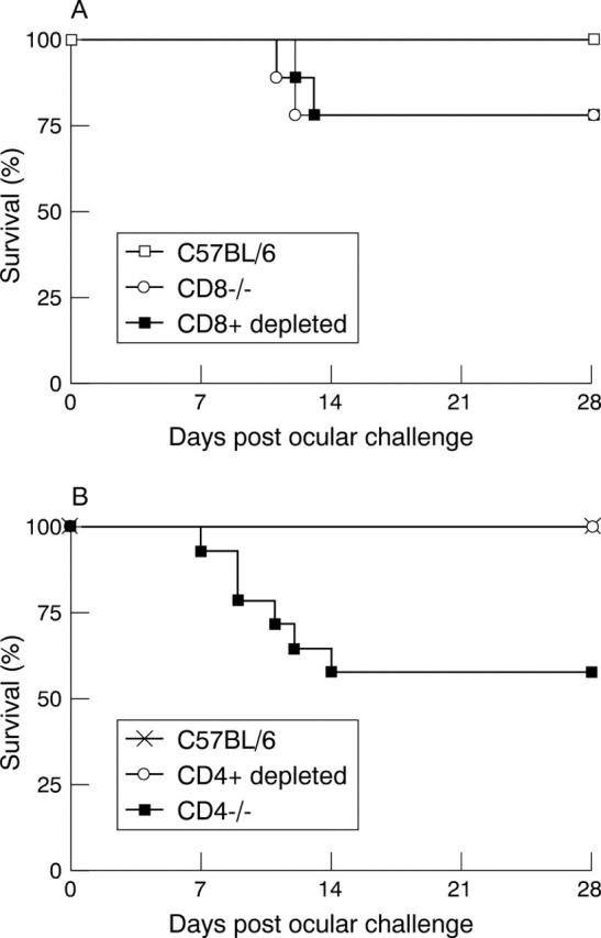
Aim: To determine the relative impact of CD4+ T cells and CD8+ T cells in protecting mice against ocular HSV-1 challenge.

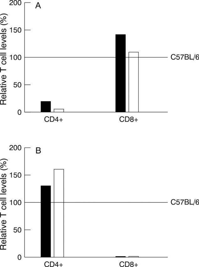
Methods: CD4+ T cell knockout mice (CD4-/- mice), CD8+ T cell knockout mice (CD8-/- mice), and mice depleted for CD4+ or CD8+ T cells by antibody (CD4+ depleted and CD8+ depleted mice), were examined for their ability to withstand HSV-1 ocular challenge. The parental mice for both knockout mice were C57BL/6J.

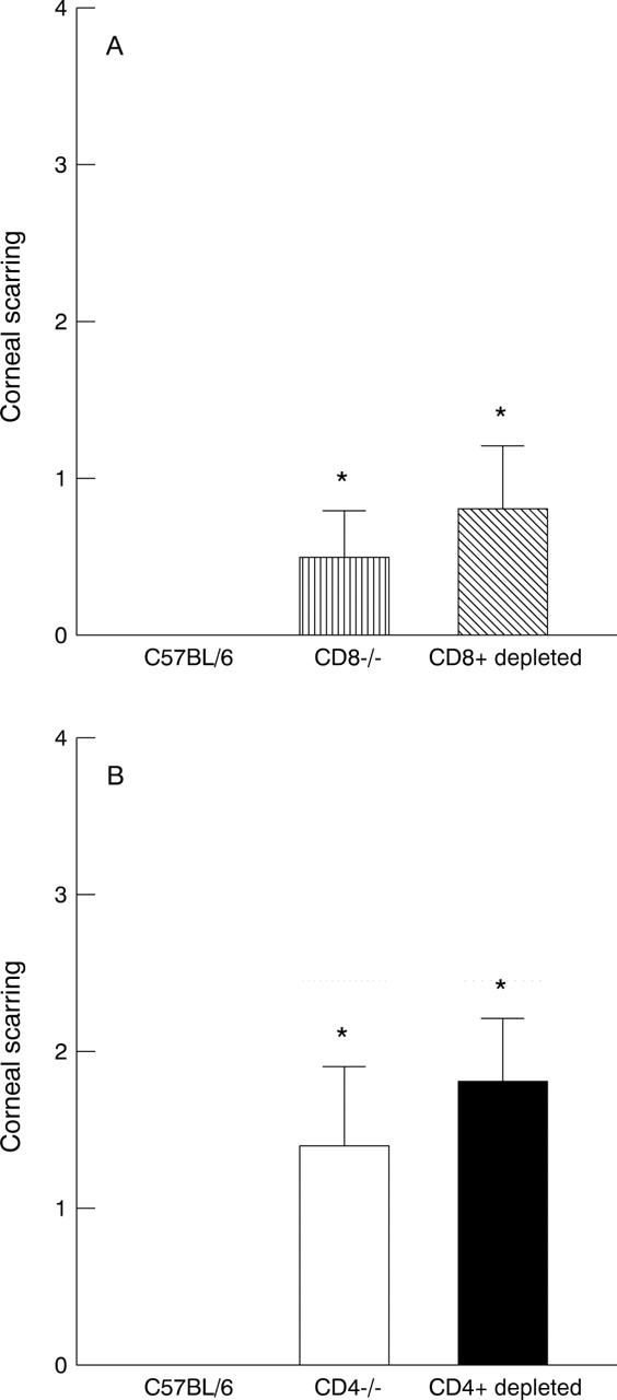
Results: These results suggest that: (1) both CD4+ deficient mice (CD4-/- and CD4+ depleted mice) and CD8+ deficient mice (CD8-/-, and CD8+ depleted mice) developed significantly more corneal scarring than their C57BL/6J parental strain; (2) the duration of virus clearance from the eyes of the CD4+ deficient mice was 4 days longer than that of the CD8+ deficient mice; and (3) the severity of corneal scarring in the CD4+ deficient mice was approximately twice that of the CD8+ deficient mice.

Conclusions: It was reported here that: (1) CD4+ and CD8+ T cells were both involved in protection against lethal ocular HSV-1 infection; and (2) CD4+ and CD8+ T cells were both involved in protection against HSV-1 induced corneal scarring.


Figures

Figure 1
Figure 1
Survival of T cell deficient mice. Mice were inoculated ocularly with 2×10⁶ pfu of McKrae as described in Materials and method. Survival was measured 28 days after ocular challenge. (A) Survival of CD8−/− and CD8+ depleted mice compared with C57BL/6J control mice after ocular infection with HSV-1. CD8−/− mice (n=10), CD8+ depleted (n=9), and C57BL/6J (n=9). (B) Survival of CD4−/− and CD4+ depleted mice compared with C57BL/6J control mice after ocular infection with HSV-1. CD4−/− mice (n=14), CD4+ depleted (n=9), and C57BL/6J (n=10).
Figure 2
Figure 2
Corneal scarring in T cell deficient mice. Corneal scarring was measured 28 days after ocular challenge as described in Materials and method in the mice shown in Figure 1 that survived ocular challenge. (A) Corneal scarring in CD8+ deficient mice; (B) corneal scarring in CD4+ deficient mice. For each bar, the corneal scarring (y axis) represents the average from 16 eyes (CD8−/− mice), 14 eyes (CD8+ depleted mice), 20 eyes (C57BL/6 mice), 16 eyes (CD4−/− mice), 18 eyes (CD4+ depleted mice), and 20 eyes (C57BL/6 mice) (SEM). *Similar to each other and significantly different from other groups (p<0.001, Student's t test).
Figure 3
Figure 3
Cytofluorimetric analysis of spleen cells from T cell deficient mice. (A) Spleens from two CD4−/− mice (solid bars) or two C57BL/6 mice depleted for CD4+ T cells with antibody (open bars) were harvested. Single cell suspensions of splenocytes were prepared, reacted with mAbs to CD4+ T cells (L3T4 mAb) and CD8+ T cells (Lyt-2 mAb) and FACS analysis was performed as described in Materials and methods. The level of CD4+CD8− T cells (CD4+) and the level of CD4−CD8+ T cells (CD8+) are shown relative to the level of these cells in untreated control C57BL/6 mice (horizontal line at 100%). Each bar represents the mean from two to four FACS analyses. (B) Spleen cells from two CD8−/− mice (solid bars) or two C57BL/6 mice depleted for CD8+ T cells with antibody (open bars) were prepared and FACS analysis performed as in (A).

References

    1. Immunity. 1996 Apr;4(4):337-47 - PubMed
    1. Invest Ophthalmol Vis Sci. 1995 Jun;36(7):1352-60 - PubMed
    1. Invest Ophthalmol Vis Sci. 1997 May;38(6):1213-21 - PubMed
    1. Immunology. 1997 Jul;91(3):430-5 - PubMed
    1. Curr Eye Res. 1997 Nov;16(11):1152-8 - PubMed

Publication types