. 2000 Apr 1;20(7):2427-38.
doi: 10.1523/JNEUROSCI.20-07-02427.2000.

Neurocan is upregulated in injured brain and in cytokine-treated astrocytes

Affiliations

Neurocan is upregulated in injured brain and in cytokine-treated astrocytes

R A Asher et al. J Neurosci. .

Abstract

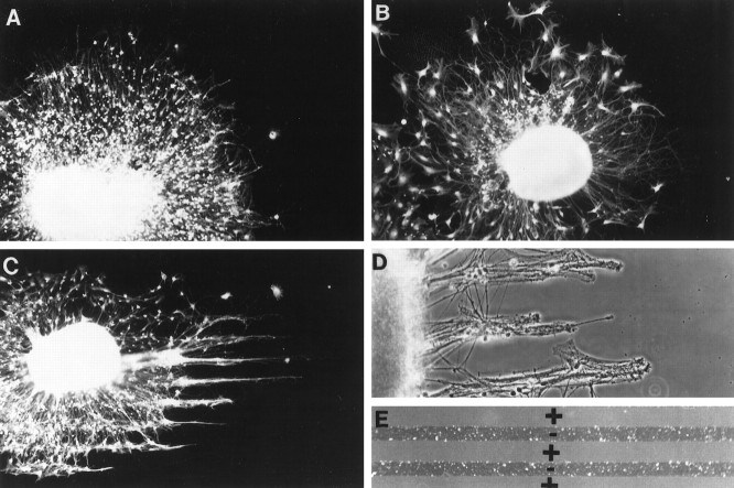
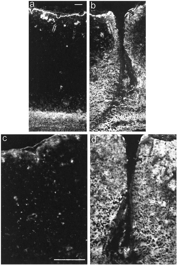
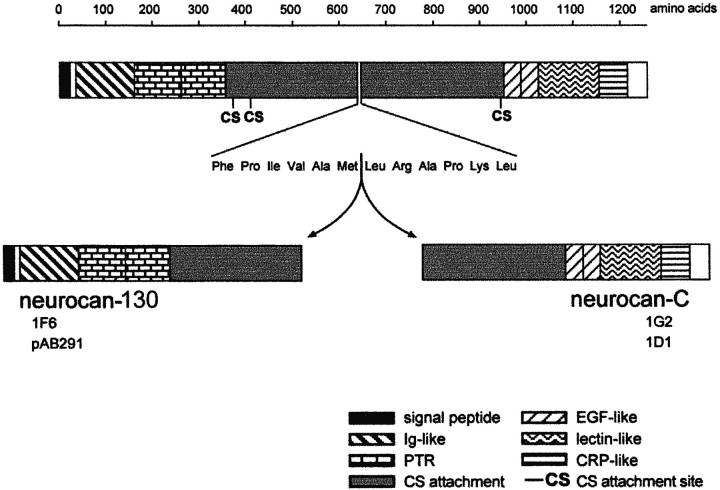
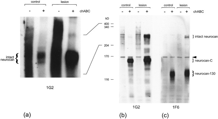
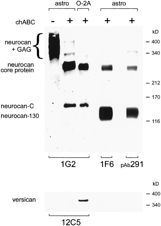
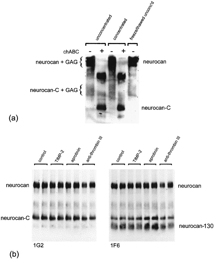
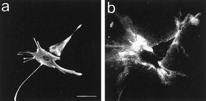
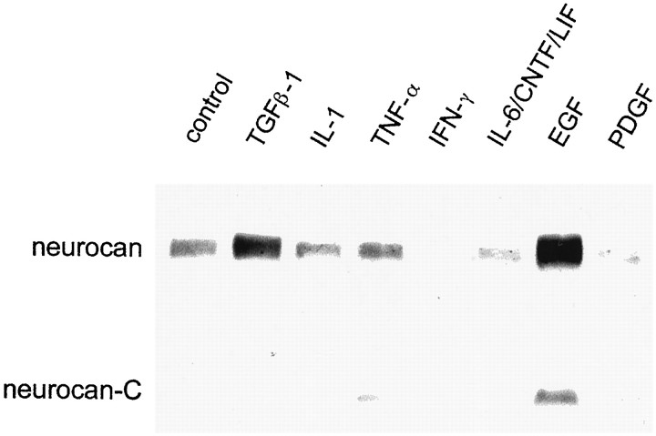
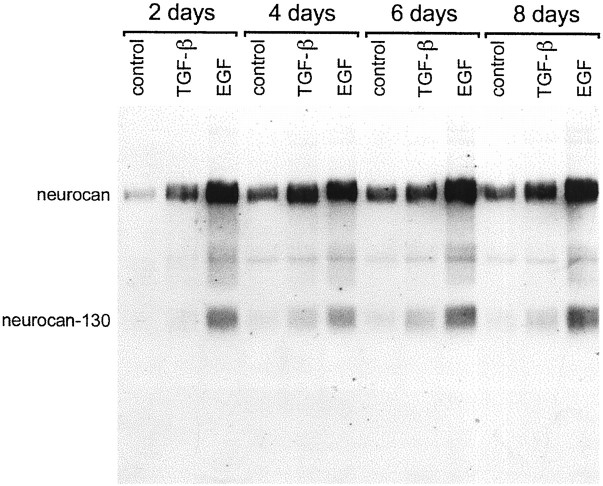
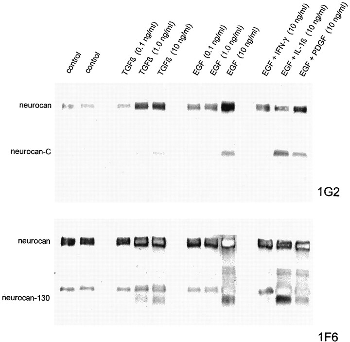
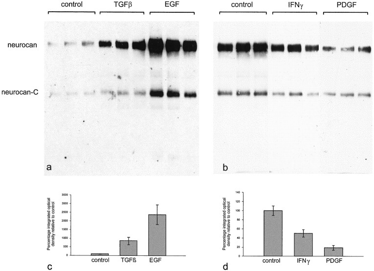
Injury to the CNS results in the formation of the glial scar, a primarily astrocytic structure that represents an obstacle to regrowing axons. Chondroitin sulfate proteoglycans (CSPG) are greatly upregulated in the glial scar, and a large body of evidence suggests that these molecules are inhibitory to axon regeneration. We show that the CSPG neurocan, which is expressed in the CNS, exerts a repulsive effect on growing cerebellar axons. Expression of neurocan was examined in the normal and damaged CNS. Frozen sections labeled with anti-neurocan monoclonal antibodies 7 d after a unilateral knife lesion to the cerebral cortex revealed an upregulation of neurocan around the lesion. Western blot analysis of extracts prepared from injured and uninjured tissue also revealed substantially more neurocan in the injured CNS. Western blot analysis revealed neurocan and the processed forms neurocan-C and neurocan-130 to be present in the conditioned medium of highly purified rat astrocytes. The amount detected was increased by transforming growth factor beta and to a greater extent by epidermal growth factor and was decreased by platelet-derived growth factor and, to a lesser extent, by interferon gamma. O-2A lineage cells were also capable of synthesizing and processing neurocan. Immunocytochemistry revealed neurocan to be deposited on the substrate around and under astrocytes but not on the cells. Astrocytes therefore lack the means to retain neurocan at the cell surface. These findings raise the possibility that neurocan interferes with axonal regeneration after CNS injury.


Figures

Fig. 1.
Fig. 1.
Neurocan is avoided by growing axons.A, A cerebellar explant growing on an L1-coated coverslip. Note the uniform halo of growing neurites. B, A cerebellar explant growing on an L1-coated coverslip with stripes of BSA (25 μg/ml). As in A, neurite outgrowth is uniform.C, A cerebellar explant growing on an L1-coated coverslip with stripes of neurocan (10 μg/ml). Axons extend in thin bundles with wider gaps between the bundles. As shown inE, the wide gaps contain the added molecules (in this case neurocan), and the narrow bands are the L1-coated substrate.D, A cortical explant growing on an L1-coated coverslip with neurocan (10 μg/ml) stripes. As in C, wide gaps separate narrow bands of outgrowth. E, Fluorescein–dextran-containing stripes (+) form wide gaps that alternate with narrow bands of uncoated substrate (). A, B,C, and E are epifluorescence illumination; D is phase-contrast optics.
Fig. 2.
Fig. 2.
Immunolocalization of neurocan in a CNS lesion. Coronal frozen sections were labeled with the anti-neurocan mAb 1G2 7 d after a knife lesion to the cerebral cortex. The images ina and b were taken with a 10× objective, and those in c and d were taken with a 20× objective. The dorsal surface of the brain isuppermost. Labeling is apparent around the lesion (b, d), which is clearly lacking on the uninjured side (a, c). Scale bar, 100 μm.
Fig. 3.
Fig. 3.
Domain structure of neurocan. The two processed fragments detected in the present experiments, neurocan-130 and neurocan-C, are thought to arise as a result of a single proteolytic cleavage on the C-terminal side of met638. Both fragments carry chondroitin sulfate. The 1G2 and 1D1 mAbs recognize epitopes in the C-terminal half of neurocan and react with neurocan-C. The 1F6 mAb and pAb291 recognize structures in the N-terminal half of neurocan and react with neurocan-130. Ig, Immunoglobulin; PTR, proteoglycan tandem repeat;CS, chondroitin sulfate; EGF, epidermal growth factor; CRP, complement regulatory protein.
Fig. 4.
Fig. 4.
Western blot analysis of neurocan expression in injured brain. Tris-buffered saline extracts were prepared from injured and uninjured cerebral cortex, and either left untreated () or treated with (+) chondroitinase ABC (chABC). The extracts were equalized for protein (15 μg in a; 8 μg in b,c), run in a 5% gel either with (b,c) or without (a) a reducing agent (0.2 m DTT), and transferred to nitrocellulose. The blots were labeled for neurocan with either the 1G2 or 1F6 mAb. Both mAbs react with intact (275 kDa) neurocan. The 1G2 mAb recognizes an epitope in the C-terminal half of neurocan and so reacts with neurocan-C, whereas 1F6 recognizes an epitope in the N-terminal half of neurocan and so reacts with neurocan-130. An upregulation of intact neurocan was clearly evident in the injured brain extracts. This difference was apparent whether or not the chondroitin sulfate was removed with chondroitinase ABC (a). The level of neurocan-C was also slightly increased (b). The band just below the 170 kDa marker (arrowhead) is nonspecific. Size determinations were made in a 5% gel by comparison of their relative mobilities with those of laminin (400 kDa), nonreduced α2-macroglobulin (340 kDa), myosin (204 kDa), α2-macroglobulin (170 kDa), and β-galactosidase (116 kDa).
Fig. 5.
Fig. 5.
Three distinct neurocan species are present in astrocyte-conditioned medium. Western blot analysis of astrocyte (astro) and O-2A lineage cells (O-2A) conditioned medium with anti-neurocan-C (1G2), anti-neurocan-130 (1F6), and anti-versican (12C5) mAbs, and a rabbit antiserum raised against neurocan-130 (pAb291). In chondroitinase-treated (+) conditioned medium, the 1G2 and 1F6 mAbs and pAb291 all recognized the same 270 kDa species (neurocan core protein). The 1G2 mAb was also reactive with a 150 kDa species (neurocan-C), whereas the 1F6 mAb and pAb291 were also reactive with a 130 kDa species (neurocan-130). Without previous chondroitinase treatment, neurocan migrated as a highMr, polydisperse species (neurocan + GAG). Neurocan core protein and neurocan-C were also detected in O-2A cell-conditioned medium. To demonstrate the specificity of these antibodies, the same samples were probed with an anti-versican mAb. No versican reactivity was detected in astrocyte-conditioned medium. A single, highMr species was seen in chondroitinase-treated O-2A cell-conditioned medium. This does not correspond to any of those recognized by the neurocan antibodies. No reactivity was seen when a mouse monoclonal IgG1 was used as the primary antibody (data not shown). Size determinations were made in a 4% gel by comparison of relative mobilities with those of the following proteins: laminin (400 kDa), nonreduced α2-macroglobulin (340 kDa), myosin (212 kDa), α2-macroglobulin (170 kDa), and β-galactosidase (116 kDa).
Fig. 6.
Fig. 6.
a, The presence of the three forms of neurocan is not an artifact of storage, concentration, or chondroitinase treatment. The same three bands are seen whether conditioned medium is analyzed before or after storing, concentrating, or chondroitinase treatment. b, Protease inhibitors do not affect neurocan processing. Astrocytes were grown in the presence of aprotinin (1.0 μg/ml), anti-thrombin III (0.1 inhibitor U/ml), or TIMP-2 (0.2 μg/ml) for 4 d. Each inhibitor was added fresh each day. Conditioned medium was concentrated and treated with chondroitinase ABC, and an equal amount of total protein (150 μg) was applied to each lane. The blot was labeled with the 1G2 mAb (left) and then relabeled with the 1F6 mAb (right). Neither serine protease (aprotinin and anti-thrombin III) nor metalloproteinase (TIMP-2) inhibitors prevented the processing of neurocan.
Fig. 7.
Fig. 7.
Astrocytes incorporate neurocan into a pericellular, substrate-bound ECM in vitro. Living astrocytes were labeled with the anti-neurocan mAb 1G2 (b), fixed in methanol, and labeled with rabbit antibodies against GFAP (a). Labeling for neurocan was seen on the substrate around GFAP-positive cells. Scale bar, 25 μm.
Fig. 8.
Fig. 8.
Cytokines influence neurocan expression in cultured astrocytes. The cells were grown for 6 d in the presence of 10 ng/ml of the cytokine, and the conditioned medium was concentrated and digested with chondroitinase ABC and an equal volume (65 μl) was applied to each lane. The blot was labeled with the anti-neurocan mAb 1G2. An increase in the amount of neurocan was seen in response to TGFβ and to a greater extent with EGF. PDGF, IFN-γ, and IL-1β brought about a decline in the amount of neurocan that was detected.
Fig. 9.
Fig. 9.
Time course of effects of TGFβ and EGF on neurocan expression in astrocytes. Cultured astrocytes were treated with TGFβ (10 ng/ml) or EGF (10 ng/ml) for 2, 4, 6, or 8 d. The conditioned medium was concentrated and treated with chondroitinase ABC, and an equal amount of protein (200 μg) was applied to each lane. The blot was labeled with the anti-neurocan mAb 1F6. TGFβ and EGF led to an increase in the amount of neurocan detected at all time points. The EGF-induced increase was at all time points greater than that of TGFβ.
Fig. 10.
Fig. 10.
Effects of the concentration of TGFβ and EGF on neurocan expression in astrocytes. Cultured astrocytes were treated with TGFβ or EGF alone (0.1, 1.0, or 10 ng/ml), or with EGF (10 ng/ml) and IFN-γ (10 ng/ml), IL-1β (10 ng/ml), or PDGF (10 ng/ml) for 4 d. The conditioned medium was concentrated and treated with chondroitinase ABC, and an equal volume (30 μl) was applied to each lane. The blot was labeled first with the anti-neurocan-C mAb 1G2 (above) and then with the anti-neurocan-130 mAb 1F6 (below). TGFβ was effective at 1.0 and 10 ng/ml but not 0.1 ng/ml, whereas EGF was only effective in bringing about an increase in neurocan expression at 10 ng/ml. IFN-γ and IL-1β appeared to override the effects of EGF. The EGF/IL-1β combination led to an increase in the amounts of neurocan-C and neurocan-130, relative to that of unprocessed neurocan.
Fig. 11.
Fig. 11.
Quantification of the effects of TGFβ, EGF, IFNγ, and PDGF on neurocan expression in astrocytes. Three flasks of astrocytes were grown in the presence of each cytokine (10 ng/ml) for 3 d. The conditioned medium was treated with chondroitinase ABC, and an equal amount of protein (50 μg in a; 200 μg in b) was applied to each lane. The blot was labeled with the anti-neurocan mAb 1G2. The amount of neurocan core protein in each lane was quantified by densitometry. TGFβ brought about a ninefold increase in the amount of neurocan detected, and EGF caused a 23-fold increase (c). PDGF reduced neurocan levels to ∼20% of control, and IFNγ brought about a 50% reduction (d). The error bars represent SEM. By Student'st test, the effects of TGFβ, EGF, and PDGF were significant with p < 0.01. The effects of IFNγ were less significant (p < 0.05).

References

    1. Asher R, Bignami A. Localization of hyaluronate in primary glial cell cultures derived from newborn rat brain. Exp Cell Res. 1991;195:401–411. - PubMed
    1. Asher R, Perides G, Vanderhaeghen J-J, Bignami A. Extracellular matrix of central nervous system white matter: demonstration of an hyaluronate-protein complex. J Neurosci Res. 1991;28:410–421. - PubMed
    1. Asher RA, Scheibe RJ, Keiser HD, Bignami A. On the existence of a cartilage-like proteoglycan and link proteins in the central nervous system. Glia. 1995;13:294–308. - PubMed
    1. Asher RA, Fidler PS, Rogers JH, Fawcett JW. TGFbeta stimulates neurocan synthesis in cultured rat astrocytes. Soc Neurosci Abstr. 1998;24:56.
    1. Asher RA, Morgenstern DA, Adcock KH, Rogers JH, Fawcett JW. Versican is up-regulated in CNS injury and is a product of O-2A lineage cells. Soc Neurosci Abstr. 1999;25:750.

Publication types

LinkOut - more resources